
Latissimus dorsi muscle
| Latissimus dorsi | |
|---|---|
trapezius muscle | |
| Identifiers | |
| Latin | musculus latissimus dorsi |
| TA98 | A04.3.01.006 |
| TA2 | 2231 |
| FMA | 13357 |
| Anatomical terms of muscle] | |
The latissimus dorsi (
The latissimus dorsi is responsible for
Due to bypassing the scapulothoracic joints and attaching directly to the spine, the actions the latissimi dorsi have on moving the arms can also influence the movement of the scapulae, such as their downward rotation during a
Structure
Variations


The number of dorsal vertebrae to which it is attached varies from four to eight; the number of costal attachments varies; muscle fibers may or may not reach the crest of the ilium.
A
A fibrous slip usually passes from the upper border of the tendon of the Latissimus dorsi, near its insertion, to the long head of the
The latissimus dorsi crosses the inferior angle of the scapula. A study found that, of 100 cadavers dissected:[6]
- 43% had "a substantial amount" of muscular fibers in the latissimus dorsi originating from the scapula.
- 36% had few or no muscular fibers, but a "soft fibrous link" between the scapula and the latissimus dorsi
- 21% had little or no connecting tissue between the two structures.
Triangles
- The lateral margin of the latissimus dorsi is separated below from the obliquus internus abdominis.
- Another triangle is situated behind the scapula. It is bounded above by the rhomboideus major. If the scapula is drawn forward by folding the arms across the chest, and the trunk bent forward, parts of the sixth and seventh ribs and the interspace between them become subcutaneous and available for auscultation. The space is therefore known as the triangle of auscultation.
- The latissimus dorsi can be remembered best for insertion as "A Miss Between Two Majors". As the latissimus dorsi inserts into the floor of the intertubercular groove of the humerus it is surrounded by two major muscles. The teres major inserts medially on the medial lip of the intertubercular groove and the pectoralis major inserts laterally onto the lateral lip.
Nerve supply
The latissimus dorsi is innervated by the sixth, seventh, and eighth cervical nerves through the thoracodorsal (long subscapular) nerve. Electromyography suggests that it consists of six groups of muscle fibres that can be independently coordinated by the central nervous system.[7]
Function
The latissimus dorsi assists in depression of the arm with the
It has a
Most latissimus dorsi exercises concurrently recruit the
Training
The power/size/strength of this muscle can be
- Vertical pulling movements such as chin-ups)
- Horizontal pulling movements such as rowing exercises
- Shoulder extension movements with straight arms such as straight-arm lat pulldowns and Pull-overs
- Deadlift
Clinical significance
Tight latissimus dorsi has been shown to be a contributor to chronic shoulder pain and chronic back pain.
The latissimus dorsi is a potential source of muscle for
Cardiac support
For heart patients with low cardiac output and who are not candidates for cardiac transplantation, a procedure called cardiomyoplasty may support the failing heart. This procedure involves wrapping the latissimus dorsi muscles around the heart and electrostimulating them in synchrony with ventricular systole.
Injury
Injuries to the latissimus dorsi are rare. They occur disproportionately in baseball pitchers. Diagnosis can be achieved by visualization of the muscle and movement testing. MRI of the shoulder girdle will confirm the diagnosis. Muscle belly injuries are treated with rehabilitation while tendon avulsion injuries can be treated surgically, or with rehab. Regardless of treatment, patients tend to return to play without any functional losses.[17]
Additional images
-
 Position of the latissimus dorsi muscle (shown in red). Animation.
 -
 Lumbar triangle
 -
 Latissimus dorsi
 -
 Clearly visible latissimus dorsi muscle of an artistic gymnast on pommel horse.
 -
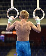 still rings.


_466.jpg)
_0857.jpg)
See also
References
![]() This article incorporates text in the public domain from the 20th edition of Gray's Anatomy (1918)
This article incorporates text in the public domain from the 20th edition of Gray's Anatomy (1918)
- ^ Kinematic Concepts for Analyzing Human Motion. In: Hall SJ. eds. Basic Biomechanics, 7e. McGraw-Hill; Accessed January 25, 2021. https://accessphysiotherapy-mhmedical-com.libaccess.lib.mcmaster.ca/content.aspx?bookid=1586§ionid=99981270
- PMID 21178031.
- S2CID 221547558.
- ^ Edwards, William E., The Musculoskeletal Anatomy of the Thorax and Brachium of an Adult Female Chimpanzee,6571st Aeromedical Research Laboratory, New Mexico, 1965. [1]
- ^ "Anatomy Atlases: Illustrated Encyclopedia of Human Anatomic Variation: Opus I: Muscular System: Alphabetical Listing of Muscles: L:Latissimus Dorsi". www.anatomyatlases.org.
- ISBN 9788847007598.
- PMID 16458022.
- S2CID 52910621.
- ISBN 9780781747806.
- ^ Arnheim, D.D., Prentice, W.E., Principles of athletic training. 9th ed. McGraw Hill, pp 570-574, 1997.
- ^ Francis, P., Applied anatomy and kinesiology, supplemental materials. KB Books., p 19-25, 1999.
- ^ Mannu, G. S., Farooq, N., Down, S., Burger, A. and Hussien, M. I. (2013), Avoiding back wound dehiscence in extended latissimus dorsi flap reconstruction. ANZ J Surg, 83: 359–364. http://onlinelibrary.wiley.com/doi/10.1111/j.1445-2197.2012.06292.x/full
- PMID 25276643.
- PMID 24419214.
- PMID 24322640.
- PMID 8983554.
- S2CID 3872258.
External links
- Anatomy figure: 01:03-08 at Human Anatomy Online, SUNY Downstate Medical Center - "Superficial layer of the extrinsic muscles of the back."
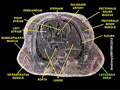
- Cross section image: pembody/body8a—Plastination Laboratory at the Medical University of Vienna