
Scaphoid fracture
| Scaphoid fracture | |
|---|---|
| Other names | Carpal scaphoid fracture, carpal navicular fracture Wrist guards[1] |
| Treatment | Not displaced: Cast[2] Displaced: Surgery[2] |
| Prognosis | Healing may take up to six months[1] |
A scaphoid fracture is a
Scaphoid fractures are most commonly caused by a fall on an outstretched hand.
The fracture may be preventable by using
It is the most commonly fractured carpal bone.[3] Males are affected more often than females.[2]
Signs and symptoms

People with scaphoid fractures generally have
Focal tenderness is usually present in one of three places: 1) volar prominence at the distal wrist for distal pole fractures; 2) anatomic snuff box for waist or midbody fractures; 3) distal to Lister's tubercle for proximal pole fractures.[4]
Complications


Avascular necrosis (AVN) is one complication of scaphoid fracture. Since the scaphoid receives its arterial supply in a retrograde fashion (i.e. from distal to proximal pole), the part proximal to the fracture is usually affected.[5]
Risk of AVN depends on the location of the fracture.
- Fractures in the proximal third have a high incidence of AVN (~30%)
- Waist fractures in the middle third is the most frequent fracture site and has moderate risk of AVN.
- Fractures in the distal third are rarely complicated by AVN.
Symptoms may include aching in the wrist, decreased range of motion of the wrist, and pain during activities such as lifting or gripping. If x-ray results show arthritis due to an old break, the treatment plan will first focus on treating the arthritis through anti-inflammatory medications and wearing a splint when an individual feels pain in the wrist. If these treatments do not help the symptoms of arthritis, steroid injections to the wrist may help alleviate pain. Should these treatments not work, surgery may be required.[6]
Mechanism

Fractures of scaphoid can occur either with direct axial compression or with hyperextension of the wrist, such as a fall on the palm on an outstretched hand. Using the Herbert classification system, there are three main types of scaphoid fractures. 10%-20% of fractures are at the proximal pole, 60%-80% are at the waist (middle), and the remainder occur at the distal pole.[4][7][8]
Diagnosis

Scaphoid fractures are often diagnosed using
-
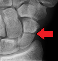
 A subtle scaphoid fracture
 -
 A more obvious scaphoid fracture on a scaphoid view X ray
 -
![Radiolucency around a 12 days old scaphoid fracture that was initially barely visible.[13]](//upload.wikimedia.org/wikipedia/commons/thumb/6/6e/Scaphoid_fracture_with_a_radiolucent_line_after_12_days.jpg/120px-Scaphoid_fracture_with_a_radiolucent_line_after_12_days.jpg) Radiolucency around a 12 days old scaphoid fracture that was initially barely visible.[13]
Radiolucency around a 12 days old scaphoid fracture that was initially barely visible.[13]


![Radiolucency around a 12 days old scaphoid fracture that was initially barely visible.[13]](/File:Scaphoid_fracture_with_a_radiolucent_line_after_12_days.jpg)
Treatment
Treatment of scaphoid fractures is guided by the location in the bone of the fracture (proximal, waist, distal), displacement (or instability) of the fracture, and patient tolerance for cast immobilization.[citation needed]
For non and minimally displaced fractures (up to 2mm) of the scaphoid waist, cast immobilisation (with surgical fixation for non-united fractures at 6 to 12 weeks) is as effective as immediate surgery fixation. This was demonstrated by the SWIFFT (Scaphoid Waist Internal Fixation for Fractures Trial) study, 439 patients were randomly allocated to either cast immobilisation or surgical fixation. There was no difference in the healing, pain and function or days off work between the two treatment groups, the cast immobilisation group had less complications and this treatment was more cost effective.
Fractures that are more proximal take longer to heal. It is expected the distal third will heal in 6 to 8 weeks, the middle third will take 8–12 weeks, and the proximal third will take 12–24 weeks.[7][8] The Scaphoid receives its blood supply primarily from lateral and distal branches of the radial artery. Blood flows from the top/distal end of the bone in a retrograde fashion down to the proximal pole; if this blood flow is disrupted by a fracture, the bone may not heal. Surgery is necessary at this point to mechanically mend the bone together.[citation needed]
Percutaneous screw fixation is recommended over an open surgical approach when it is possible to achieve acceptable bone alignment closed as minimal incisions can preserves the palmar ligament complex and local vasculature, and help avoid soft tissue complications. This surgery includes screwing the scaphoid bone back together at the most perpendicular angle possible to promote quicker and stronger healing of the bone. Internal fixation can be done dorsally with a percutaneous incision and arthroscopic assistance [17] or via a minimal open dorsal approach,[16] or via a volar approach in which case slight excavation of the edge of the trapezium bone may be necessary to reach the scaphoid as 80% of this bone is covered with articular cartilage, which makes it difficult to gain access to the scaphoid.[18]
Prognosis
A non-union (
In the aftermath, 90% of non-operated individuals return to sports, with 88% reaching their previous level. Among those who underwent surgery, the rate of returning to sports is 98%, and 96% return to their previous level. The average time observed for resuming sports is 14 weeks for non-operated individuals and 7 weeks for those who had surgery.[20]
Epidemiology
Fractures of the scaphoid are common in young males.[21] They are less common in children and older adults because the distal radius is weaker contributor to the wrist and more likely to fracture in these age groups.[7] Scaphoid fractures account for 50%-80% of carpal injuries.[8]
Terminology
These are also called navicular fractures (the scaphoid also being called the carpal navicular), although this can be confused with the navicular bone in the foot.[citation needed]
References
- ^ a b c d e f g h i "Scaphoid Fracture of the Wrist". AAOS. March 2016. Archived from the original on 24 September 2017. Retrieved 12 October 2017.
- ^ PMID 15368727.
- PMID 26051761.
- ^ a b c deWeber K. "Scaphoid fractures". UpToDate.com. Archived from the original on 2013-10-29.
- PMID 27252968.
- ^ Jones B (2010). "Scaphoid Fracture of the Wrist". Ortho Info. American Academy of Orthopedic Surgeons. Archived from the original on December 7, 2015. Retrieved November 30, 2015.
- ^ OCLC 706805938.
- ^ OCLC 960851324.
- ^ Jones J. "Scaphoid series". Radiology Reference Article. Radiopaedia.org. Retrieved 2021-08-30.
- PMID 29027844.
- ^ "BestBets: Magnetic resonance imaging of suspected scaphoid fractures". Archived from the original on 2010-06-16.
- PMID 19756904.
- ^ S2CID 221014238."
- ^ PMID 33109331.
- ^ S2CID 33543861.
- ^ Slade JF 3rd, Gutow AP, Geissler WB. Percutaneous internal fixation of scaphoid fractures via an arthroscopically assisted dorsal approach. J Bone Joint Surg Am. 2002;84-A Suppl 2:21-36. doi:10.2106/00004623-200200002-00003
- ^ Kastelec M. "Percutaneous Screw Fixation". AO Foundation. Archived from the original on December 8, 2015. Retrieved November 30, 2015.
- PMID 29027844.
- PMID 30788227.
- ISBN 9781282950023.
External links
| Authority control databases: National |
|---|