
Spermatocytic tumor
| Spermatocytic tumor | |
|---|---|
| Other names | Spermatocytic tumour |
 | |
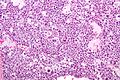
| Micrograph of a spermatocytic tumor. H&E stain. | |
| Specialty | Pathology, urology |
Spermatocytic tumor, previously called spermatocytic seminoma, is a
The name of the tumour comes from the similarity (under the
Signs and symptoms
Spermatocytic tumor is a rare tumour, making up only one to two percent of all testicular germ cell tumours. Men presenting with this tumour are generally 50 to 60 years old, and its occurrence is rare in men under 30 years old. Most present with slow, painless testicular enlargement, which may involve both testes.[1]
Diagnosis
Spermatocytic tumors are diagnosed based on tissue from orchiectomy (or partial orchiectomy), done for a lesion suspicious for cancer on medical imaging.[citation needed]
The
Histologic appearance
- small cells with a large μm),
- medium-sized cells with prominent nucleoli(15-18 μm) and,
- large cells (50-100 μm).
The cells are generally packed into nodules, and have a loose, sheet-like arrangement that is commonly interrupted by interstitial oedema. Unlike classical seminoma, fibrous septation and lymphocytic infiltrates are not seen. Cells undergoing mitosis are common, as are cells undergoing apoptosis.[1]
Intratubular growth of spermatocytic tumor can be seen, however there is no
Rarely, spermatocytic tumors may show sarcomatoid differentiation, most commonly as undifferentiated spindled cells intermingled within the typical-appearing spermatocytic tumor cells. Rhabdomyosarcomatous differentiation has also been described.[1]
-
 Micrograph of a spermatocytic tumor. H&E stain.

Relation to seminoma
Spermatocytic tumor is not considered a subtype of
Treatment
Unlike classical seminoma, spermatocytic tumors rarely metastasise, so radical orchidectomy alone is sufficient treatment, and retroperitoneal lymph node dissection and adjuvant chemotherapy or radiotherapy are generally not required.[1]
References
- ^ ISBN 978-0-7817-7942-5
- ISBN 0-7216-0187-1.
- PMID 3583416.
External links
- Spermatocytic seminoma micrograph - webpathology.com.