
Sublingual gland
| Sublingual gland | |
|---|---|
Sublingual artery (branch of lingual artery of external carotid artery) | |
| Nerve | Submandibular ganglion |
| Lymph | Submandibular lymph nodes |
| Identifiers | |
| Latin | glandula sublingualis |
| MeSH | D013361 |
| TA98 | A05.1.02.008 |
| TA2 | 2807 |
| FMA | 59791 |
| Anatomical terminology] | |
The sublingual gland (glandula sublingualis) is a seromucous polystomatic exocrine gland. Located underneath the oral diaphragm (diaphragma oris), the sublingual gland is the smallest and most diffuse of the three major salivary glands of the oral cavity, with the other two being the submandibular and parotid. The sublingual gland provides approximately 3-5% of the total salivary volume.[1][2]
Structure
They lie anterior and superior to the submandibular gland and inferior and lateral to the tongue, as well as beneath the mucous membrane of the floor of the mouth. They are bound laterally by the bone of the mandible and inferolaterally by the mylohyoid muscle. The glands can be felt behind each mandibular canine. Placing one index finger within the mouth and the fingertips of the opposite hand outside it, the compressed gland is manually palpated between the inner and outer fingers.[clarification needed][3]
The sublingual gland is constituted by 1 major duct and approximately 20 small excretory ducts, with the latter often being referred to as ducts of Rivinus. The sublingual gland consists mostly of mucous acini capped with 
Microanatomy
Blood supply
The gland receives its blood supply from the sublingual and submental arteries.[4] Lymph from the sublingual salivary gland drains into the submandibular lymph nodes.[3]
Nerve supply
The
Development
The sublingual salivary glands appear in the eighth week of prenatal development, two weeks later than the other two major salivary glands. They develop from epithelial buds in the sulcus surrounding the sublingual folds on the floor of the mouth, lateral to the developing submandibular gland. These buds branch and form into cords that canalize to form the sublingual ducts associated with the gland. The rounded terminal ends of the cords form acini.[1]
Clinical significance
Ranulas are the most common pathologic lesion associated with the sublingual glands.[5]
Additional images
-
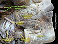
 Imaging showing the sublingual glands and surrounding structures.
 -
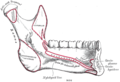 Mandible. Inner surface. Side view.
 -
 Sublingual gland
 -
 Sublingual gland
 -
 Sublingual gland


References
- ^ a b c Illustrated Dental Embryology, Histology, and Anatomy, Bath-Balogh and Fehrenbach, Elsevier, 2011, page 136-137
- ^ "Submandibular Gland: Location, Function and Complications".
- ^ a b c Illustrated Anatomy of the Head and Neck, Fehrenbach and Herring, Elsevier, 2012, p. 156
- ^ a b Ten Cate's Oral Histology, Nanci, Elsevier, 2013, page 255
- ISBN 9780323049030.
External links
- Anatomy photo:34:st-0701 at the SUNY Downstate Medical Center - "Oral Cavity: Glands"
- cranialnerves at The Anatomy Lesson by Wesley Norman (Georgetown University) (VII)
- "Anatomy diagram: 25420.000-1". Roche Lexicon - illustrated navigator. Elsevier. Archived from the original on 2015-02-26.
- Salivary gland infections from Medline Plus
- Salivary gland cancer from American Cancer Society