
Lingual tonsils
| Lingual tonsils | |
|---|---|
 Tongue | |
| Details | |
| System | Immune system (Lymphatic system) |
| Identifiers | |
| Latin | tonsilla lingualis |
| TA98 | A05.1.04.022 |
| TA2 | 2830 |
| FMA | 54836 |
| Anatomical terminology | |
The lingual tonsils are a collection of
Structure
Microanatomy
Lingual tonsils are covered externally by stratified squamous nonkeratinized epithelium that invaginates inward forming crypts. Beneath the epithelium is a layer of lymphoid nodules containing lymphocytes. Mucous glands located at the root of tongue are drained through several ducts into the crypt of lingual tonsils.[2][3] Secretions of these mucous glands keep the crypt clean and free of any debris.
Blood supply
Lingual tonsils are located on posterior aspect of tongue which is supplied through:[1]
- Lingual artery, branch of external carotid artery
- Tonsillar branch of facial artery
- Ascending and descending palatine arteries
- Ascending pharyngeal branch of external carotid artery
Nerve supply
Lingual tonsils are innervated by tonsillar nerves from the tonsilar plexus, formed by the glossopharyngeal and vagus nerves.[1]
Function

Like other lymphatic tissues, the function of lingual tonsils is to prevent infections. These tonsils contain B and T lymphocytes which get activated when harmful bacteria and viruses come in contact with tonsils. B lymphocytes kill pathogens by producing antibodies against them, while T lymphocytes directly kill them releasing cytotoxic substances or indirectly by stimulating other cells of the immune system.[2][3][4]
Clinical significance
Cancer
Sleep apnea
Enlarged or hypertrophic lingual tonsils have the potential to cause or exacerbate sleep apnea.[6]
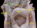
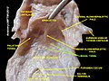
Additional images
-
 Lingual tonsil
 -
 Lingual tonsil
 -
 Lingual tonsils


![]() This article incorporates text in the public domain from page 1138 of the 20th edition of Gray's Anatomy (1918)
This article incorporates text in the public domain from page 1138 of the 20th edition of Gray's Anatomy (1918)
External links
- Pictures at usc.edu(Registration required)
- Anatomy Atlases – Microscopic Anatomy, plate 09.163
- Histology image: 09802loa – Histology Learning System at Boston University
- MedEd at Loyola histo/HistoImages/hl6-27.jpg (labeled as 'lymphoid tissue')]
- Lingual Tonsil