User:Tvarghes/sandbox
Lateral Globus Pallidus

The lateral globus pallidus (or external globus pallidus, GPe) combines with the
The lateral globus pallidus contains
GPe is particular in comparison to the other elements of the basal ganglia by the fact that it does not work as an output base of the basal ganglia but as a main regulator of the basal ganglia system.
Function

Indirect Striatopallidal Pathway
The
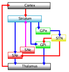
The Indirect Striatopallidal Pathway, which contains the lateral globus pallidus and the subthalamic nucleus, functions to modulate the effects of the Direct Striatopallidal Pathway. The lateral globus pallidus acts as a inhibitory "control device", adjusting subthalamic nucleus neuronal activity via GABAergic output. [3].
When movement adjustment is required, striatal inhibitory
This
Related Pathology
Lateral globus pallidus dysfunction has been observed in the following conditions:
Medial Globus Pallidus

The medial globus pallidus (or internal, GPi) combines with the
The medial globus pallidus contains
Function

The medial globus pallidus acts to tonically inhibit the ventral lateral nucleus and ventral anterior nucleus of the thalamus. As these two nuclei are needed for movement planning, this inhibition restricts movement initiation and prevents unwanted movements.
The Direct Striatopallidal Pathway
The medial globus pallidus receives inhibitory GABAergic signals from the striatum when a movement requirement is signaled from the cerebral cortex. As the medial globus pallidus is one of the direct output centers of the basal ganglia, this causes disinihibtion of the thalamus, increasing overall ease of initiating and maintaining movement. As this pathway only contains one synapse (from the striatum to the medial globus pallidus), it is considered a direct pathway.[5]
The Direct Striatopallidal Pathway is modulated by stimulation of the medial globus pallidus by the
Clinical Significance
Pathology
Dysfunction of the medial globus pallidus has been correlated to the following conditions:
Deep brain stimulation
Deep brain stimulation (DBS) is a treatment by which regulated electrical pulses are sent to specifically targeted areas, and has been used on the medial globus pallidus to treat a variety of medical conditions.
DBS has been applied to patients with Tardive Dyskinesia, and the majority saw more than 50% improvement in symptoms[9]. Tourette Syndrome patients have also benefited from this treatment, showing over 50% improvement in tic severity (compulsive disabling motor tics are symptoms of Tourette patients)[8]. The medial globus pallidus is also considered a "highly effective target for neuromodulation" when using deep brain stimulation on Parkinson's Disease patients[6].
References
- ^ PMID 24416002.)
{{cite journal}}: CS1 maint: unflagged free DOI (link - .
- ^ .
- PMID 26841063.
- PMID 26785520.
- ^ PMID 19398341.
- ISBN 9783662463437.
- ^ PMID 23206487.
- ^ PMID 23099106.
