
Cerebral cortex
| Cerebral cortex | |
|---|---|
 | |
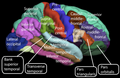
 Motor and sensory areas of the cerebral cortex | |
| Details | |
| Part of | Cerebrum |
| Identifiers | |
| Latin | cortex cerebri |
| MeSH | D002540 |
| NeuroNames | 39 |
| NeuroLex ID | birnlex_1494 |
| TA98 | A14.1.09.003 A14.1.09.301 |
| TA2 | 5527, 5528 |
| FMA | 61830 |
| Anatomical terms of neuroanatomy | |
The cerebral
The six-layered neocortex makes up approximately 90% of the cortex, with the allocortex making up the remainder.[3] The cortex is divided into left and right parts by the longitudinal fissure, which separates the two cerebral hemispheres that are joined beneath the cortex by the corpus callosum. In most mammals, apart from small mammals that have small brains, the cerebral cortex is folded, providing a greater surface area in the confined volume of the cranium. Apart from minimising brain and cranial volume, cortical folding is crucial for the brain circuitry and its functional organisation.[4] In mammals with small brains, there is no folding and the cortex is smooth.[5][6]
A fold or ridge in the cortex is termed a gyrus (plural gyri) and a groove is termed a sulcus (plural sulci). These surface convolutions appear during fetal development and continue to mature after birth through the process of gyrification. In the human brain, the majority of the cerebral cortex is not visible from the outside, but buried in the sulci.[7] The major sulci and gyri mark the divisions of the cerebrum into the lobes of the brain. The four major lobes are the frontal, parietal, occipital and temporal lobes. Other lobes are the limbic lobe, and the insular cortex often referred to as the insular lobe.
There are between 14 and 16 billion
Structure

The cerebral cortex is the outer covering of the surfaces of the cerebral hemispheres and is folded into peaks called gyri, and grooves called sulci. In the human brain, it is between 2 and 3-4 mm. thick,[8] and makes up 40% of the brain's mass.[2] 90% of the cerebral cortex is the six-layered neocortex whilst the other 10% made up of three/four-layered allocortex.[2] There are between 14 and 16 billion neurons in the cortex,[2] and these are organized radially in cortical columns, and minicolumns, in the horizontally organized layers of the cortex.[9][10]
The neocortex is separable into different regions of cortex known in the plural as cortices, and include the motor cortex and visual cortex. About two thirds of the cortical surface is buried in the sulci and the insular cortex is completely hidden. The cortex is thickest over the top of a gyrus and thinnest at the bottom of a sulcus.[11]
Folds
The cerebral cortex is folded in a way that allows a large surface area of neural tissue to fit within the confines of the neurocranium. When unfolded in the human, each hemispheric cortex has a total surface area of about 0.12 square metres (1.3 sq ft).[12] The folding is inward away from the surface of the brain, and is also present on the medial surface of each hemisphere within the longitudinal fissure. Most mammals have a cerebral cortex that is convoluted with the peaks known as gyri and the troughs or grooves known as sulci. Some small mammals including some small rodents have smooth cerebral surfaces without gyrification.[6]
Lobes
The larger sulci and gyri mark the divisions of the cortex of the cerebrum into the lobes of the brain.[8] There are four main lobes: the frontal lobe, parietal lobe, temporal lobe, and occipital lobe. The insular cortex is often included as the insular lobe.[13] The limbic lobe is a rim of cortex on the medial side of each hemisphere and is also often included.[14] There are also three lobules of the brain described: the paracentral lobule, the superior parietal lobule, and the inferior parietal lobule.
Thickness
For species of mammals, larger brains (in absolute terms, not just in relation to body size) tend to have thicker cortices.[15] The smallest mammals, such as shrews, have a neocortical thickness of about 0.5 mm; the ones with the largest brains, such as humans and fin whales, have thicknesses of 2–4 mm.[2][8] There is an approximately logarithmic relationship between brain weight and cortical thickness.[15] Magnetic resonance imaging of the brain (MRI) makes it possible to get a measure for the thickness of the human cerebral cortex and relate it to other measures. The thickness of different cortical areas varies but in general, sensory cortex is thinner than motor cortex.[16] One study has found some positive association between the cortical thickness and intelligence.[17] Another study has found that the
Layers of neocortex


The neocortex is formed of six layers, numbered I to VI, from the outermost layer I – near to the pia mater, to the innermost layer VI – near to the underlying white matter. Each cortical layer has a characteristic distribution of different neurons and their connections with other cortical and subcortical regions. There are direct connections between different cortical areas and indirect connections via the thalamus.
One of the clearest examples of cortical layering is the line of Gennari in the primary visual cortex. This is a band of whiter tissue that can be observed with the naked eye in the calcarine sulcus of the occipital lobe. The line of Gennari is composed of axons bringing visual information from the thalamus into layer IV of the visual cortex.
Staining cross-sections of the cortex to reveal the position of neuronal cell bodies and the intracortical axon tracts allowed neuroanatomists in the early 20th century to produce a detailed description of the laminar structure of the cortex in different species. The work of Korbinian Brodmann (1909) established that the mammalian neocortex is consistently divided into six layers.
Layer I
Layer I is the molecular layer of cerebral cortex|molecular layer, and contains few scattered neurons, including
Layer II
Layer II, the external granular layer, contains small pyramidal neurons and numerous stellate neurons.
Layer III
Layer III, the external pyramidal layer, contains predominantly small and medium-size pyramidal neurons, as well as non-pyramidal neurons with vertically oriented intracortical axons; layers I through III are the main target of interhemispheric corticocortical afferents, and layer III is the principal source of corticocortical efferents.
Layer IV
Layer IV, the
Layer V
Layer V, the internal pyramidal layer, contains large pyramidal neurons. Axons from these leave the cortex and connect with subcortical structures including the
Layer VI
Layer VI, the polymorphic layer or multiform layer, contains few large pyramidal neurons and many small spindle-like pyramidal and multiform neurons; layer VI sends
Columns
The cortical layers are not simply stacked one over the other; there exist characteristic connections between different layers and neuronal types, which span all the thickness of the cortex. These cortical microcircuits are grouped into cortical columns and minicolumns.[31] It has been proposed that the minicolumns are the basic functional units of the cortex.[32] In 1957, Vernon Mountcastle showed that the functional properties of the cortex change abruptly between laterally adjacent points; however, they are continuous in the direction perpendicular to the surface. Later works have provided evidence of the presence of functionally distinct cortical columns in the visual cortex (Hubel and Wiesel, 1959),[33] auditory cortex, and associative cortex.
Cortical areas that lack a layer IV are called agranular. Cortical areas that have only a rudimentary layer IV are called dysgranular.[34] Information processing within each layer is determined by different temporal dynamics with that in layers II/III having a slow 2 Hz oscillation while that in layer V has a fast 10–15 Hz oscillation.[35]
Types of cortex
Based on the differences in laminar organization the cerebral cortex can be classified into two types, the large area of neocortex which has six cell layers, and the much smaller area of allocortex that has three or four layers:[3]
- The neocortex is also known as the isocortex or neopallium and is the part of the mature cerebral cortex with six distinct layers. Examples of neocortical areas include the granular primary visual cortex. The neocortex has two subtypes, the true isocortex and the proisocortexwhich is a transitional region between the isocortex and the regions of the periallocortex.
- The allocortex is the part of the cerebral cortex with three or four layers, and has three subtypes, the olfactory cortex and the hippocampus.
There is a transitional area between the neocortex and the allocortex called the paralimbic cortex, where layers 2, 3 and 4 are merged. This area incorporates the proisocortex of the neocortex and the periallocortex of the allocortex. In addition, the cerebral cortex may be classified into four lobes: the frontal lobe, temporal lobe, the parietal lobe, and the occipital lobe, named from their overlying bones of the skull.
Blood supply and drainage

Blood supply to the cerebral cortex is part of the cerebral circulation. Cerebral arteries supply the blood that perfuses the cerebrum. This arterial blood carries oxygen, glucose, and other nutrients to the cortex. Cerebral veins drain the deoxygenated blood, and metabolic wastes including carbon dioxide, back to the heart.
The main arteries supplying the cortex are the anterior cerebral artery, the middle cerebral artery, and the posterior cerebral artery. The anterior cerebral artery supplies the anterior portions of the brain, including most of the frontal lobe. The middle cerebral artery supplies the parietal lobes, temporal lobes, and parts of the occipital lobes. The middle cerebral artery splits into two branches to supply the left and right hemisphere, where they branch further. The posterior cerebral artery supplies the occipital lobes.
The circle of Willis is the main blood system that deals with blood supply in the cerebrum and cerebral cortex.

Development
The
Neural tube
The cerebral cortex develops from the most anterior part, the forebrain region, of the
Cortical neuron development
Cortical neurons are generated within the ventricular zone, next to the ventricles. At first, this zone contains neural stem cells, that transition to radial glial cells–progenitor cells, which divide to produce glial cells and neurons.[40]
Radial glia

The cerebral cortex is composed of a heterogenous population of cells that give rise to different cell types. The majority of these cells are derived from radial glia migration that form the different cell types of the neocortex and it is a period associated with an increase in neurogenesis. Similarly, the process of neurogenesis regulates lamination to form the different layers of the cortex. During this process there is an increase in the restriction of cell fate that begins with earlier progenitors giving rise to any cell type in the cortex and later progenitors giving rise only to neurons of superficial layers. This differential cell fate creates an inside-out topography in the cortex with younger neurons in superficial layers and older neurons in deeper layers. In addition, laminar neurons are stopped in S or G2 phase in order to give a fine distinction between the different cortical layers. Laminar differentiation is not fully complete until after birth since during development laminar neurons are still sensitive to extrinsic signals and environmental cues.[41]
Although the majority of the cells that compose the cortex are derived locally from radial glia there is a subset population of neurons that
At birth there are very few dendrites present on the cortical neuron's cell body, and the axon is undeveloped. During the first year of life the dendrites become dramatically increased in number, such that they can accommodate up to a hundred thousand synaptic connections with other neurons. The axon can develop to extend a long way from the cell body.[45]
Asymmetric division
The first divisions of the progenitor cells are symmetric, which duplicates the total number of progenitor cells at each
Layer organization

The
Cortical patterning

The map of functional cortical areas, which include primary motor and visual cortex, originates from a '
Rapid expansion of the cortical surface area is regulated by the amount of self-renewal of
Evolution
Of all the different brain regions, the cerebral cortex shows the largest evolutionary variation and has evolved most recently.[6] In contrast to the highly conserved circuitry of the medulla oblongata, for example, which serves critical functions such as regulation of heart and respiration rates, many areas of the cerebral cortex are not strictly necessary for survival. Thus, the evolution of the cerebral cortex has seen the advent and modification of new functional areas—particularly association areas that do not directly receive input from outside the cortex.[6]
A key theory of cortical evolution is embodied in the radial unit hypothesis and related protomap hypothesis, first proposed by Rakic.[62] This theory states that new cortical areas are formed by the addition of new radial units, which is accomplished at the stem cell level. The protomap hypothesis states that the cellular and molecular identity and characteristics of neurons in each cortical area are specified by cortical stem cells, known as radial glial cells, in a primordial map. This map is controlled by secreted signaling proteins and downstream transcription factors.[63][64][65]
Function

Connections
The cerebral cortex is connected to various subcortical structures such as the thalamus and the basal ganglia, sending information to them along efferent connections and receiving information from them via afferent connections. Most sensory information is routed to the cerebral cortex via the thalamus. Olfactory information, however, passes through the olfactory bulb to the olfactory cortex (piriform cortex). The majority of connections are from one area of the cortex to another, rather than from subcortical areas; Braitenberg and Schüz (1998) claim that in primary sensory areas, at the cortical level where the input fibers terminate, up to 20% of the synapses are supplied by extracortical afferents but that in other areas and other layers the percentage is likely to be much lower.[66]
Cortical areas
The whole of the cerebral cortex was divided into 52 different areas in an early presentation by Korbinian Brodmann. These areas known as Brodmann areas, are based on their cytoarchitecture but also relate to various functions. An example is Brodmann area 17 which is the primary visual cortex.
In more general terms the cortex is typically described as comprising three parts: sensory, motor, and association areas.
Sensory areas

The sensory areas are the cortical areas that receive and process information from the
Motor areas
The motor areas are located in both hemispheres of the cortex. The motor areas are very closely related to the control of voluntary movements, especially fine fragmented movements performed by the hand. The right half of the motor area controls the left side of the body, and vice versa.
Two areas of the cortex are commonly referred to as motor:
- Primary motor cortex, which executes voluntary movements [citation needed]
- Supplementary motor areas and premotor cortex, which select voluntary movements. [citation needed]
In addition, motor functions have been described for:
- Posterior parietal cortex, which guides voluntary movements in space
- Dorsolateral prefrontal cortex, which decides which voluntary movements to make according to higher-order instructions, rules, and self-generated thoughts.
Just underneath the cerebral cortex are interconnected subcortical masses of grey matter called
Association areas

The association areas are the parts of the cerebral cortex that do not belong to the primary regions. They function to produce a meaningful perceptual experience of the world, enable us to interact effectively, and support abstract thinking and language. The parietal, temporal, and occipital lobes – all located in the posterior part of the cortex – integrate sensory information and information stored in memory. The frontal lobe or prefrontal association complex is involved in planning actions and movement, as well as abstract thought. Globally, the association areas are organized as distributed networks.[69] Each network connects areas distributed across widely spaced regions of the cortex. Distinct networks are positioned adjacent to one another yielding a complex series of interwoven networks. The specific organization of the association networks is debated with evidence for interactions, hierarchical relationships, and competition between networks.
In humans, association networks are particularly important to language function. In the past it was theorized that language abilities are localized in Broca's area in areas of the left inferior frontal gyrus, BA44 and BA45, for language expression and in Wernicke's area BA22, for language reception. However, the processes of language expression and reception have been shown to occur in areas other than just those structures around the lateral sulcus, including the frontal lobe, basal ganglia, cerebellum, and pons.[70]
Clinical significance

Neurodegenerative diseases such as Alzheimer's disease, show as a marker, an atrophy of the grey matter of the cerebral cortex.[72]
Other diseases of the central nervous system include neurological disorders such as epilepsy, movement disorders, and different types of aphasia (difficulties in speech expression or comprehension).
The developing fetus is susceptible to a range of environmental factors that can cause birth defects and problems in later development. Maternal alcohol consumption for example can cause fetal alcohol spectrum disorder.[73] Other factors that can cause neurodevelopment disorders are toxicants such as drugs, and exposure to radiation as from X-rays. Infections can also affect the development of the cortex. A viral infection is one of the causes of lissencephaly, which results in a smooth cortex without gyrification.
A type of electrocorticography called cortical stimulation mapping is an invasive procedure that involves placing electrodes directly onto the exposed brain in order to localise the functions of specific areas of the cortex. It is used in clinical and therapeutic applications including pre-surgical mapping.[74]
Genes associated with cortical disorders
There are a number of genetic mutations that can cause a wide range of genetic disorders of the cerebral cortex, including microcephaly, schizencephaly and types of lissencephaly.[75] Chromosome abnormalities can also result causing a number of neurodevelopmental disorders such as fragile X syndrome and Rett syndrome.
Mutations in
History
In 1909, Korbinian Brodmann distinguished different areas of the neocortex based on cytoarchitectural difference and divided the cerebral cortex into 52 regions.[78]
Other animals
The cerebral cortex is derived from the
In avian brains, evidence suggests the avian pallium's neuroarchitecture to be reminiscent of the mammalian cerebral cortex.[79] The avian pallium has also been suggested to be an equivalent neural basis for consciousness.[80][81]
Until recently no counterpart to the cerebral cortex had been recognized in invertebrates. However, a study published in the journal Cell in 2010, based on gene expression profiles, reported strong affinities between the cerebral cortex and the mushroom bodies of the ragworm Platynereis dumerilii.[82] Mushroom bodies are structures in the brains of many types of worms and arthropods that are known to play important roles in learning and memory; the genetic evidence indicates a common evolutionary origin, and therefore indicates that the origins of the earliest precursors of the cerebral cortex date back to the Precambrian era.
Additional images
-
 Lateral surface of the human cerebral cortex
 -
 Medial surface of the human cerebral cortex
 -
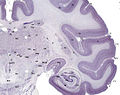 Tissue slice from the brain of an adult macaque monkey. The cerebral cortex is depicted in dark violet.


See also
References
- ^ "cerebral mantle". TheFreeDictionary.com.
- ^ ISBN 9780071222075.
- ^ ISBN 978-1-61779-778-1.
- ^ PMID 17580069.
- PMID 27056680.
- ^ PMID 19763105.
- ISBN 978-0838577011.
- ^ ISBN 9780387977775.
- PMID 26359774.
Functional columns were first defined in the cortex by Mountcastle (1957), who proposed the columnar hypothesis, which states that the cortex is composed of discrete, modular columns of neurons, characterized by a consistent connectivity profile.
- PMID 27932026.
- ISBN 978-0683014556.
- PMID 18267953.
- PMID 22230626.
- ISBN 9780470646083.
- ^ ISBN 978-3-540-56013-5.
- PMID 12946467.
- PMID 17118969.
- PMID 18025393.
- ^ Catharine Paddock (2007-11-20). "Migraine Sufferers Have Thicker Brain Cortex". Medical News Today. Archived from the original on 2008-05-11.
- PMID 21911412.
- PMID 11099442.
- ^ "Scientists identify a new kind of human brain cell". Allen Institute. 27 August 2018.
- PMID 10600995.
- S2CID 143409890.
- PMID 17553419.
- S2CID 4015532.
- PMID 19188274.
- ^ S2CID 17846130.
- ^ Creutzfeldt, O. 1995. Cortex Cerebri. Springer-Verlag.
- ^ PMID 19447861.
- S2CID 36706690.
- PMID 9153131.
- PMID 14403679.
- ^ S.M. Dombrowski, C.C. Hilgetag, and H. Barbas. Quantitative Architecture Distinguishes Prefrontal Cortical Systems in the Rhesus Monkey Archived 2008-08-29 at the Wayback Machine.Cereb. Cortex 11: 975–988. "...they either lack (agranular) or have only a rudimentary granular layer IV (dysgranular)."
- PMID 19805197.
- PMID 24373884.
- ISBN 9780199678143.
- PMID 10498281.
- ISBN 0-443-06583-7
- S2CID 3041502.
- S2CID 893478.
- ^ ISBN 978-0-12-374539-2.
- PMID 19763105.
- S2CID 31900267.
- ISBN 9780878932504.
- PMID 16014714.
- PMID 3291116.
- PMID 22549586.
- S2CID 21371568.
- S2CID 10881759.
- PMID 11466432.
- PMID 3291116.
- S2CID 14807054.
- S2CID 6533589.
- PMID 10764649.
- PMID 22031906.
- PMID 21414909.
- PMID 21729779.
- PMID 23622239.
- PMID 27214567.
- PMID 23804101.
- PMID 3291116.
- S2CID 14807054.
- PMID 10764649.
- S2CID 12282525.
- ^ Braitenberg, V and Schüz, A 1998. "Cortex: Statistics and Geometry of Neuronal Connectivity. Second thoroughly revised edition" New York: Springer-Verlag
- ^ Saladin, Kenneth. Anatomy and Physiology: The Unity of Form and Function, 5th Ed. New York: McGraw-Hill Companies Inc., 2010. Print.
- ^ Dorland's Medical Dictionary for Health Consumers, 2008.
- PMID 21653723.
- PMID 11117622.
- S2CID 210196036.
- PMID 34670843.
- PMID 16738372.
- PMID 22702484.
- ^ PMID 15148137.
- ^ "EMX2 empty spiracles homeobox 2 [Homo sapiens (human)] – Gene – NCBI". www.ncbi.nlm.nih.gov.
- PMID 26879631.
- ^ )
- ISSN 0036-8075.
- S2CID 221881862.
- S2CID 221882004.
- S2CID 917306.
External links
- hier-20 at NeuroNames
- Stained brain slice images which include the "cerebral cortex" at the BrainMaps project
- "The primary visual cortex", Webvision: Comprehensive article about the structure and function of the primary visual cortex.
- "Basic cell types", Webvision: Image of the basic cell types of the monkey cerebral cortex.
- Cerebral Cortex – Cell Centered Database