The clavicle, collarbone, or keybone is a slender, S-shaped
abducted. The clavicle is the most commonly fractured bone. It can easily be fractured by impacts to the shoulder from the force of falling on outstretched arms or by a direct hit.[3]
The rounded medial region (sternal region) of the shaft has a long curve laterally and anteriorly along two-thirds of the entire shaft. The flattened lateral region (acromial region) of the shaft has an even larger posterior curve to articulate with the acromion of the scapula. The medial region is the longest clavicular region as it takes up two-thirds of the entire shaft. The lateral region is both the widest clavicular region and thinnest clavicular region. The lateral end has a rough inferior surface that bears a ridge, the trapezoid line, and a slight rounded projection, the conoid tubercle (above the coracoid process). These surface features are attachment sites for muscles and ligaments of the shoulder.
It can be divided into three parts: medial end, lateral end, and shaft.
Medial end
The medial end is also known as the sternal end. It is quadrangular and articulates with the clavicular notch of the manubrium of the sternum to form the sternoclavicular joint. The articular surface extends to the inferior aspect for articulation with the first costal cartilage.
Lateral end
The lateral end is also known as the acromial end. It is flat from above downward. It bears a facet that articulates with the shoulder to form the acromioclavicular joint. The area surrounding the joint gives an attachment to the joint capsule. The anterior border is concave forward and the posterior border is convex backward.
Shaft
The shaft is divided into two main regions, the medial region, and the lateral region. The medial region is also known as the sternal region, it is the longest clavicular region as it takes up two-thirds of the entire shaft. The lateral region is also known as the acromial region, it is both the widest clavicular region and thinnest clavicular region.
3D model of the clavicle
Lateral region of the shaft
The lateral region of the shaft has two borders and two surfaces.
the anterior border is concave forward and gives origin to the deltoid muscle.
the posterior border is convex and gives attachment to the
trapezius muscle
.
the inferior surface has a ridge called the trapezoid line and a tubercle; the conoid tubercle for attachment with the trapezoid and the conoid ligament, part of the coracoclavicular ligament that serves to connect the collarbone with the coracoid process of the scapula.
Parts of clavicle
Development
The collarbone is the first bone to begin the process of
ossification centres, one medial and one lateral, which fuse later on. The compact forms as the layer of fascia
covering the bone stimulate the ossification of adjacent tissue. The resulting compact bone is known as a periosteal collar.
derived from elements originally attached to the skull.
Variation
The shape of the clavicle varies more than most other long bones. It is occasionally pierced by a branch of the
supraclavicular nerve. In males the clavicle is usually longer and larger than in females. A study measuring 748 males and 252 females saw a difference in collarbone length between age groups 18–20 and 21–25 of about 6 and 5 mm (0.24 and 0.20 in) for males and females respectively.[7]
The left clavicle is usually longer and weaker than the right clavicle.[6][8]
The levator claviculae muscle, present in 2–3% of people, originates on the transverse processes of the upper cervical vertebrae and is inserted in the lateral half of the clavicle.
It serves as a rigid support from which the scapula and free limb are suspended; an arrangement that keeps the upper limb away from the thorax so that the arm has maximum range of movement. Acting as a flexible, crane-like strut, it allows the scapula to move freely on the thoracic wall.
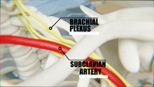
Relation of Brachial Plexus with the ClavicleCovering the cervicoaxillary canal, it protects the neurovascular bundle that supplies the upper limb.
Transmits physical impacts from the upper limb to the axial skeleton.
Muscle
Muscles and ligaments that attach to the collarbone include:
A vertical line drawn from the mid-clavicle called the
mid-clavicular line is used as a reference in describing cardiac apex beat during medical examination. It is also useful for evaluating an enlarged liver, and for locating the gallbladder which is between the mid-clavicular line and the transpyloric plane
Clavicle fractures (colloquially, a broken collarbone) occur as a result of injury or trauma. The most common type of fractures occur when a person falls horizontally on the shoulder or with an outstretched hand. A direct hit to the collarbone will also cause a break. In most cases, the direct hit occurs from the lateral side towards the medial side of the bone. The most common site of fracture is the junction between the two curvatures of the bone, which is the weakest point.[9] This results in the sternocleidomastoid muscle lifting the medial aspect superiorly, which can result in perforation of the overlying skin.
Other animals
The clavicle first appears as part of the skeleton in primitive
cartilaginous fish and in the vast majority of living bony fish, including all of the teleosts.[10]
placental mammals. In many mammals, the clavicles are also reduced, or even absent, to allow the scapula greater freedom of motion, which may be useful in fast-running animals.[10]
theropod dinosaurs to form a furcula, which is the equivalent to a wishbone.[12]
In birds, the clavicles and interclavicle have fused to form a single Y-shaped bone, the furcula or "wishbone" which evolved from the clavicles found in coelurosaurian theropods.


 Position of collarbone (shown in red). Animation.
 Shape of collarbone (left). Animation.
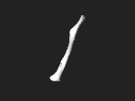 3D image
 Pectoral girdle—front
 Diagram of the human shoulder joint, front view
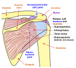 Diagram of the human shoulder joint, back view
 Muscles of the neck. Anterior view.
 Clavicle


