
Costal cartilage
| Costal cartilage | |
|---|---|
interchondral articulations. Anterior view. | |
| Details | |
| Identifiers | |
| Latin | cartilagines costales |
| Greek | costo condrio |
| MeSH | D066186 |
| TA98 | A02.3.01.005 |
| TA2 | 1140, 1139 |
| FMA | 7591 |
| Anatomical terminology] | |
The costal cartilages are bars of hyaline cartilage[1] that serve to prolong the ribs forward and contribute to the elasticity of the walls of the thorax. Costal cartilage is only found at the anterior ends of the ribs, providing medial extension.
Differences from Ribs 1-12
The first seven pairs are connected with the
Like the ribs, the costal cartilages vary in their length, breadth, and direction. They increase in length from the first to the seventh, then gradually decrease to the twelfth.
Their breadth, as well as that of the intervals between them, diminishes from the first to the last. They are broad at their attachments to the ribs, and taper toward their sternal extremities, excepting the first two, which are of the same breadth throughout, and the sixth, seventh, and eighth, which are enlarged where their margins are in contact.
They also vary in direction: the first descends a little to the sternum, the second is horizontal, the third ascends slightly, while the others are angular, following the course of the ribs for a short distance, and then ascending to the sternum or preceding cartilage.
Structure
Each costal cartilage presents two surfaces, two borders, and two extremities.
Surfaces
The anterior surface is convex, and looks forward and upward: that of the first gives attachment to the costoclavicular ligament and the subclavius muscle; those of the first six or seven at their sternal ends, to the pectoralis major. The others are covered by, and give partial attachment to, some of the flat muscles of the abdomen.
The posterior surface is concave, and directed backward and downward; that of the first gives attachment to the
Borders
Of the two borders the superior is concave, the inferior convex; they afford attachment to the
The inferior borders of the sixth, seventh, eighth, and ninth cartilages present heel-like projections at the points of greatest convexity. These projections carry smooth oblong facets which articulate with facets on slight projections from the upper borders of the seventh, eighth, ninth, and tenth cartilages, respectively.
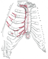
Interchondral articulations
The interchondral articulations are the
Extremities
The lateral end of each cartilage is continuous with the osseous tissue of the rib to which it belongs.
The medial end of the first is continuous with the sternum; the medial ends of the six succeeding ones are rounded and are received into shallow concavities on the lateral margins of the sternum.
The medial ends of the eighth, ninth, and tenth costal cartilages are pointed, and are connected each with the cartilage immediately above.
Those of the eleventh and twelfth are pointed and free.
Clinical significance
In old age, the costal cartilages are prone to superficial ossification, particularly in women with age of 50 years and over.[3]
In costochondritis and Tietze syndrome, inflammation of the costal cartilage occurs.[4] This is a common cause of chest pain.[5]
Severe trauma may lead to fracture of the costal cartilage.[6] Such injuries often go unnoticed during x-ray scans, but can be diagnosed with CT scans.[6] Surgery is typically used to fix the costal cartilage back onto either the rib or sternum.[6]
Costal cartilage may be
Additional images
-
 Position of the costal cartilages (shown in red). Animation.
 -
 Anterior surface ofsternumand costal cartilages.


See also
- Costochondral joint
- Costochondritis
- Human rib cage
- Rib
References
![]() This article incorporates text in the public domain from page 127 of the 20th edition of Gray's Anatomy (1918)
This article incorporates text in the public domain from page 127 of the 20th edition of Gray's Anatomy (1918)