
Dermoid cyst
This article needs additional citations for verification. (April 2015) |
| Dermoid cyst | |
|---|---|
Gynecology |
A dermoid cyst is a
tissue.As dermoid cysts grow slowly and contain mature tissue, this type of cystic teratoma is nearly always
Location
Due to its classification, a dermoid cyst can occur wherever a teratoma can occur.
Vaginal and ovarian dermoid cysts
Ovaries normally grow cyst-like structures called follicles each month. Once an egg is released from its follicle during ovulation, follicles typically deflate. Sometimes fluid accumulates inside the follicle, forming a simple (containing only fluid) cyst.[2] The majority of these functional cysts resolve spontaneously.[citation needed]
While all
-
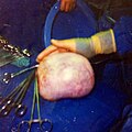 Large ovarian cyst
 -
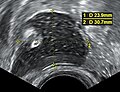 Dermoid cyst in vaginal ultrasonography
 -
 A complex cyst due to a dermoid as seen on ultrasound
 -
 A complex cyst due to a dermoid as seen on CT. Arrow points to bone or teeth.


Mark.png)
Periorbital dermoid cysts
Dermoid cysts can appear in young children, often near the lateral aspect of the eyebrow (right part of the right eyebrow or left part of the left eyebrow). Depending on the perceived amount of risk, these are sometimes excised or simply kept under observation.
An inflammatory reaction can occur if a dermoid cyst is disrupted, and the cyst can recur if it is not completely excised. Sometimes complete excision is not practical if the cyst is in a dumbbell configuration, whereby it extends through a suture line in the skull.
If dermoid cysts appear on the medial aspect, the possibility of an encephalocele becomes greater and should be considered among the differential diagnoses.
Other areas where a dermoid cyst may appear are the brain, scrotum and the pharynx.
Dermoid cysts develop during pregnancy. They occur when skin cells and things like hair, sweat glands, oil glands or fatty tissue get trapped in the skin as a baby grows in the womb. Dermoid cysts are present at birth (congenital) and are common. It can be months or years before a dermoid cyst is noticed on a child because the cysts grow slowly.
Dermoid cyst symptoms are minor and the cysts are usually painless. They are not harmful to a child's health. If they become infected, the infection must be treated and the cyst should be removed. It is easier to remove cysts and prevent scars if the cyst is removed before it gets infected.
Spinal dermoid cysts
Spinal dermoid cysts are benign ectopic growths thought to be a consequence of embryology errors during neural tube closure. Their reported incidence is extremely rare, accounting for less than 1% of intramedullary spinal cord tumours. It has been proposed that a possible 180 cases of spinal dermoid tumours have been identified over the past century in the literature.[8][9]
Dermoid cysts more often involve the
Various hypotheses have been advanced to explain the pathogenesis of spinal dermoids, the origin of which may be acquired or congenital.
- Acquired or iatrogenic dermoids may arise from the implantation of epidermal tissue into the subdural space i.e. spinal cutaneous inclusion, during needle puncture (e.g. lumbar puncture) or during surgical procedures on closure of a dysraphic malformation.[9][10]
- Congenital dermoids, however, are thought to arise from cells whose position is correct but which fail to
Spinal abnormalities, e.g. intramedullary dermoid cysts may arise more frequently in the lumbosacral region (quite often at the level of the conus medullaris) and may be seen with other congenital anomalies of the spine including posterior spina bifida occulta as identified by the neuroradiological analysis.[8][11]
Diagnosis
Differential diagnosis
A small dermoid cyst on the
Treatment
Treatment for dermoid cyst is complete surgical removal, preferably in one piece and without any spillage of cyst contents.
The association of dermoid cysts with pregnancy has been increasingly reported. They usually present the dilemma of weighing the risks of surgery and anesthesia versus the risks of untreated adnexal mass. Most references state that it is more feasible to treat bilateral dermoid cysts of the ovaries discovered during pregnancy if they grow beyond 6 cm in diameter.
See also
- pilonidal cyst
- Proliferating trichilemmal cyst
- List of cutaneous conditions
References
- ISBN 0-07-138076-0.
- ^ "Ovarian Cysts". Mayo Clinic. Mayo Foundation for Medical Education and Research.
- S2CID 30041721.
- ^ "Medical Definition of Dermoid cyst of the ovary". MedicineNet.
- ISBN 9780781765275. Retrieved 22 February 2018 – via Google Books.
- ^ "Tumours of the Vagina; Chapter Six" (PDF). International Agency for Research on Cancer, World Health Organization. pp. 291–311. Archived from the original (PDF) on 2015-09-08.
- ^ "Vulva and Vagina tumors: an overview". atlasgeneticsoncology.org. Retrieved 2018-02-22.
- ^ S2CID 6453067.
- ^ S2CID 22201164.
- ^ PMID 5937643.
- ^ S2CID 7264952.
- ^ Maki A (November 16, 2006). "Maki:Jones returns to say goodbye". The Globe and Mail.