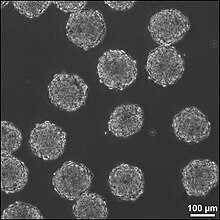
Embryoid body

Embryoid bodies (EBs) are three-dimensional aggregates formed by pluripotent
EBs are differentiation of human embryonic stem cells into embryoid bodies comprising the three embryonic germ layers. They mimic the characteristics seen in early-stage embryos. They are often used as a model system to conduct research on various aspects of developmental biology. They can also contribute to research focused on tissue engineering and regenerative medicine.
Background
The pluripotent cell types that comprise embryoid bodies include
In contrast to monolayer cultures, however, the spheroid structures that are formed when ESCs aggregate enables the non-adherent culture of EBs in suspension, making EB cultures inherently scalable, which is useful for bioprocessing approaches, whereby large yields of cells can be produced for potential clinical applications.
Formation
EBs are formed by the
Formation of EBs can also be more precisely controlled by the inoculation of known cell densities within single drops (10-20 µL) suspended from the lid of a Petri dish, known as hanging drops.[21] While this method enables control of EB size by altering the number of cells per drop, the formation of hanging drops is labor-intensive and not easily amenable to scalable cultures. Additionally, the media can not be easily exchanged within the traditional hanging drop format, necessitating the transfer of hanging drops into bulk suspension cultures after 2–3 days of formation, whereby individual EBs tend to agglomerate. Recently, new technologies have been developed to enable media exchange within a modified hanging drop format.[28] In addition, technologies have also been developed to physically separate cells by forced aggregation of ESCs within individual wells or confined on adhesive substrates,[29][30][31][32] which enables increased throughput, controlled formation of EBs. Ultimately, the methods used for EB formation may impact the heterogeneity of EB populations, in terms of aggregation kinetics, EB size and yield, as well as differentiation trajectories.[31][33][34]
Differentiation within EBs
Within the context of ESC
As a result of the three-dimensional EB structure, complex morphogenesis occurs during EB differentiation, including the appearance of both epithelial- and mesenchymal-like cell populations, as well as the appearance of markers associated with the
Parallels with embryonic development
Much of the research central to embryonic stem cell differentiation and morphogenesis is derived from studies in developmental biology and mammalian embryogenesis.
In addition, advancements of EB culture resulted in the development of
Challenges to directing differentiation
In contrast to the differentiation of ESCs in monolayer cultures, whereby the addition of soluble morphogens and the extracellular microenvironment can be precisely and homogeneously controlled, the three-dimensional structure of EBs poses challenges to directed differentiation.
See also
- Brain organoid
- Gastruloid
- Induced stem cells
- Pluripotency
References
- PMID 6950406.
- S2CID 4256553.
- PMID 7544005.
- PMID 9804556.
- PMID 16589125.
- S2CID 4260518.
- PMID 10985386.
- S2CID 1565219.
- S2CID 8531539.
- S2CID 86129154.
- PMID 18691744.
- PMID 10859025.
- ^ PMID 3897439.
- PMID 15153605.
- ^ PMID 18295582.
- ^ PMID 19198003.
- PMID 18260134.
- PMID 20017699.
- ^ PMID 17609152.
- PMID 8898231.
- ^ PMID 16683985.
- PMID 12930726.
- S2CID 4346252.
- S2CID 11484871.
- S2CID 8213725.
- PMID 20406903.
- S2CID 25461651.
- S2CID 35415772.
- PMID 17653344.
- PMID 16884768.
- ^ PMID 19805103.
- PMID 18270562.
- PMID 19193130.
- PMID 18583540.
- PMID 10949929.
- PMID 12382948.
- PMID 6365932.
- PMID 11352941.
- PMID 7585945.
- PMID 9885251.
- PMID 10974008.
- PMID 14696356.
- PMID 1893864.
- PMID 18550259.
- PMID 18371422.
- ^ PMID 18983966.
- PMID 20026669.
- S2CID 22010083.
- S2CID 4421136.
- PMID 10410899.
- PMID 7521282.
- PMID 12972006.
- S2CID 20596850.
- PMID 16289026.
- ^ bioRxiv 10.1101/104539.
- ^ bioRxiv 10.1101/051722.
- ^ PMID 26650833.
- PMID 25371360.
- PMID 25371361.
- ^ PMID 21491967.
- ^ PMID 19162317.
- PMID 18793799.
- S2CID 42754482.
- PMID 20864164.
- PMID 22079776.
- PMID 22076329.
- PMID 20001738.
- PMID 18724832.
