Leptomeningeal collateral circulation
| Leptomeningeal collateral circulation | |
|---|---|
 Arterial supply of the brain | |
| Details | |
| Location | around the brain |
| Function | small connections (anastamoses) between the areas supplied by the major arteries of the brain. |
| Anatomical terminology | |
The leptomeningeal collateral circulation (also known as leptomeningeal anastomoses or pial collaterals) is a network of small blood vessels in the brain that connects branches of the middle, anterior and posterior cerebral arteries (MCA, ACA, and PCA),[1] with variation in its precise anatomy between individuals.[2] During a stroke, leptomeningeal collateral vessels allow limited blood flow when other, larger blood vessels provide inadequate blood supply to a part of the brain.[3]
Structure


Leptomeningeal collaterals lie within the leptomeninges, the two deep layers of the meninges called the pia mater and the arachnoid mater.[4] Their diameter has been measured at approximately 300 micrometers,[5] but there is variability between individuals in the size, quantity and location of these vessels, and between either hemisphere within the same subject.[6]
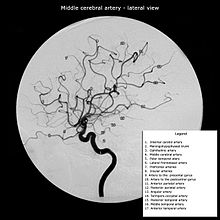
Inter-territorial end to end anastomoses exist between branches of the anterior cerebral artery and middle cerebral artery, the posterior cerebral artery and middle cerebral artery, the anterior cerebral artery and posterior cerebral artery, and the right and left anterior cerebral arteries.[7][8][9][10] Intra-territorial anastamoses connect adjacent arterial branches within the same arterial territory (between two branches of the same middle cerebral artery, for example).[5]
| Inter-territorial leptomeningeal anastamoses relative to branches of the middle cerebral artery[5] | |
|---|---|
| Supplying the Frontal Lobe | |
| Prefrontal arteries | No anastamoses observed |
| Orbito-frontal (lateral frontobasal) artery | Anterior and inferior frontal arteries (branches of the anterior cerebral artery) |
| Precentral (pre-rolandic) artery | Posterior inferior frontal artery (a branch of the anterior cerebral artery) |
| Central (rolandic) artery | Paracentral artery (a branch of the anterior cerebral artery) |
| Supplying the Parietal Lobe | |
| Anterior parietal artery | Precuneal artery (a branch of the anterior cerebral artery) |
| Posterior parietal artery | No anastamoses observed |
| Angular artery | Parieto-occipital artery (a branch of the posterior cerebral artery) |
| Temporo-occipital | No anastamoses observed |
| Supplying the Temporal Lobe | |
| Posterior temporal artery | No anastamoses observed |
| Middle temporal artery | No anastamoses observed |
| Anterior temporal artery | Anterior temporal artery (a branch of the posterior cerebral artery) |
| Temporopolar artery | No anastamoses observed |
Inter-territorial leptomeningeal anastamoses between the posterior cerebral artery and anterior cerebral artery have been observed between the parieto-occipital branch of the posterior cerebral artery, and the precuneal branch or the posterior pericallosal branch of the anterior cerebral artery.[1]
Inter-territorial leptomeningeal anastamoses between the right and left anterior cerebral arteries have been observed between the right and left pericallosal arteries and the right and left callosal marginal arteries. Anastamoses have also been observed between precuneal branches originating from the middle portion of the pericallosal artery, or from the posterior portion of the callosal marginal branch of one side joining the opposite paracentral branch.[1]
There is anatomical variation in collateral circulation from person to person, and as we age, collateral vessels decrease in diameter and number.[2]
Function
Leptomeningeal collateral vessels allow limited cerebral blood flow and brain tissue perfusion when the brain receives insufficient blood supply through an artery, via a series of anastomotic connections between cerebral arteries.[3]
Clinical significance
Stroke

During an ischaemic stroke, blood flow through a cerebral artery is compromised. This frequently causes substantial injury to the area of the brain supplied by the artery, but not all of this territory is necessarily affected. A post mortem study of middle cerebral artery strokes demonstrated that the area of brain injury was often smaller than the total area supplied by the middle cerebral artery. Leptomeningeal collateral vessels from the anterior cerebral artery and posterior cerebral artery appeared to allow for perfusion of some brain tissue to persist, partially compensating for the loss of the major vessel.[6] This compensatory effect is however usually inadequate to maintain a normal blood supply.[11]
Therapies that attempt to optimize leptomeningeal collateral circulation appear to improve outcomes following acute ischaemic stroke.[2]
MRI and CT brain imaging is used to determine the severity of a stroke, and help guide treatment. Fluid attenuated inversion recovery (FLAIR) vascular hyperintensity (FVH) is a radiographic marker seen on brain imaging in acute ischaemic stroke. FVH can be used as a proxy for slow leptomeningeal collateral blood flow, and may help reveal which areas of brain tissue are potentially salvageable.[12]
Alzheimer’s disease
The age-related changes that can be seen in leptomeningeal vessels over time appear to be accelerated by Alzheimer's disease, according to mouse models conducted in 2018.[13]
Intracranial haemorrhage
A 2016 study compared patients awaiting carotid artery stenting for unilateral atherosclerotic plaques. Those with leptomeningeal collaterals evident on cranial angiography had a higher incidence of intracranial haemorrhage (ICH) after stenting. The authors argued that the presence of such collaterals on imaging should be considered a risk factor for ICH in patients where carotid stenting is otherwise indicated.[14]
History

The term 'leptomeningeal' derives from the Greek word leptos (λεπτός) meaning thin, in reference to the appearance of the pia mater and arachnoid mater.
Descriptions of leptomeningeal collateral vessels are found in Thomas Willis’ Cerebri Anatome (1664).[15][16] German physician Otto Heubner first demonstrated their presence in his 1874 work Die luetische Erkrankung Der Hirnaterien.[17] He injected the middle cerebral artery, anterior cerebral artery and posterior cerebral artery in turn, in an attempt to establish the territories these arteries supply. Even when other anastomoses from the circle of Willis were blocked off, the whole cerebral arterial tree could be filled.[1] Later study in the 1950s and 60s by H.M. Vander Eecken and R.D. Adams provided a comprehensive review of the anatomy of the leptomeningeal collateral circulation.[6]
The concept of the ischaemic penumbra, where brain tissue shows capacity to recover if perfusion is quickly restored, was defined in 1981 by Astrup et al. Persistent blood flow through leptomeningeal vessels is a key part of this recovery.[18]
Other animals
Haemodynamic studies of leptomeningeal collaterals have been conducted in primates.[19] Leptomeningeal circulation has been observed in mice and rats during experiments to assess changes associated with disease and ageing in these vessels.[20]
References
- ^ PMID 14576375.
- ^ S2CID 206189480.
- ^ PMID 22518231.
- PMID 20099639.
- ^ PMID 25285093.
- ^ S2CID 33711931.
- ^ Cipolla, Marilyn J. (2009). Anatomy and Ultrastructure. Morgan & Claypool Life Sciences.
- PMID 8938760.
- PMID 21885720.
- )
- PMID 9763379.
- PMID 27659851.
- PMID 30519973.
- S2CID 24415856.
- PMID 16528199.
- ^ Willis, Thomas (1664). Cerebri anatome : cui accessit Nervorum descriptio et usus /. Londini: Typis Tho. Roycroft, impensis Jo. Martyn & Ja. Allestry ...
- ^ "Die luetische Erkrankung der Hirnarterien". Wellcome Library. Retrieved 2019-03-10.
- PMID 19365124.
- PMID 4969977.
- PMID 30239233.
