
Middle pharyngeal constrictor muscle
| Middle pharyngeal constrictor muscle | |
|---|---|
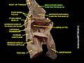
pharynx and cheek (middle pharyngeal constrictor muscle labeled as constrictor pharyngis medius at center left) | |
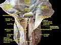
 Muscles of the pharynx, viewed from behind, together with the associated vessels and nerves (middle pharyngeal constrictor muscle labeled as Mid. constr. at center) | |
| Details | |
| Origin | Hyoid bone |
| Insertion | Pharyngeal raphe |
| Artery | Ascending pharyngeal artery |
| Nerve | Pharyngeal plexus of vagus nerve |
| Actions | Swallowing |
| Identifiers | |
| Latin | musculus constrictor pharyngis medius |
| TA98 | A05.3.01.108 |
| TA2 | 2184 |
| FMA | 46622 |
| Anatomical terms of muscle] | |
The middle pharyngeal constrictor is a fan-shaped
The middle pharyngeal constrictor originates from the
Structure
The middle pharyngeal constrictor is a sheet-like, fan-shaped muscle.[1]
The muscle's fibers diverge from their origin: the more inferior fibres descend deep to the
Origin
Two parts of the middle pharyngeal constrictor muscle are distinguished according to its sites of origin:
- Ceratopharyngeal part - arises (the entire superior margin of) the greater cornu of the hyoid bone.[1]
- Chondropharyngeal part - arises from the lesser cornu of the hyoid bone, and (the inferior portion of) the stylohyoid ligament. The chondropharyngeal part represents the muscle's anterior origin.[1]
Insertion
The muscle inserts (posteriorly) into the pharyngeal raphe,[1] blending with its contralateral partner at the midline.[citation needed]
Innervation
Similarly to the superior and inferior pharyngeal constrictor muscles, it is innervated by a branch of the vagus nerve through the pharyngeal plexus.[citation needed]
Actions/movements
The contraction of the muscle constricts the middle portion of the pharynx.[1]
Function
The muscle contracts during swallowing:[1] as soon as the bolus of food is received in the pharynx, the elevator muscles relax, the pharynx descends, and the constrictors contract upon the bolus, and convey it downward towards the esophagus.[2][3]
They also have respiratory mechanical effects.[4]
Additional images
-
 Hyoid bone. Anterior surface. Enlarged.
 -
 Muscles of the neck. Lateral view.
 -
 Middle pharyngeal constrictor muscle
 -
 Middle pharyngeal constrictor muscle
 -
 Deep dissection of larynx, pharynx and tongue seen from behind
 -
 Deep dissection of larynx, pharynx and tongue seen from behind
 -
 Deep dissection of larynx, pharynx and tongue seen from behind


References
![]() This article incorporates text in the public domain from page 1143 of the 20th edition of Gray's Anatomy (1918)
This article incorporates text in the public domain from page 1143 of the 20th edition of Gray's Anatomy (1918)
Further reading
- Its role in speech: Hamaker, Ronald C.; Blom, Eric D. (2003). "Botulinum Neurotoxin for Pharyngeal Constrictor Muscle Spasm in Tracheoesophageal Voice Restoration". The Laryngoscope. 113 (9): 1479–1482. S2CID 12251825.
- Its role in Hyoid bone syndrome: Ernest, Edwin A.; Salter, E. George (1991). "Hyoid bone syndrome: A degenerative injury of the middle pharyngeal constrictor muscle with photomicroscopic evidence of insertion tendinosis". The Journal of Prosthetic Dentistry. 66 (1): 78–83. PMID 1941681.
External links
- lesson8 at The Anatomy Lesson by Wesley Norman (Georgetown University) (latpharyngealitems3)