
Mylohyoid nerve
| Mylohyoid nerve | |
|---|---|
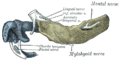
Mandibular division of the trigeminal nerve. (Label for mylohyoid nerve is at bottom center.) | |
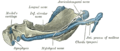
 Mandibular division of trifacial nerve, seen from the middle line. The small figure is an enlarged view of the otic ganglion. (Label "to mylohyoid" at bottom left.) | |
| Details | |
| From | inferior alveolar nerve |
| Innervates | mylohyoid muscle, anterior belly of digastric muscle |
| Identifiers | |
| Latin | nervus mylohyoideus |
| TA98 | A14.2.01.090 |
| TA2 | 6275 |
| FMA | 53247 |
| Anatomical terms of neuroanatomy] | |
The mylohyoid nerve (or nerve to mylohyoid) is a
Structure
Origin
The mylohyoid nerve is a mixed (motor-sensory)[1] branch of the inferior alveolar nerve (which is a branch of the mandibular nerve (CN V3) that is itself a branch of the trigeminal nerve (CN V)).[2][1] It arises just before it enters the mandibular foramen.[1]
Course
It pierces the
Distribution
Motor
The mylohyoid nerve supplies the
Sensory
It provides sensory innervation to the skin of the centre of the submental area.[4] It may also provide some sensory innervation to the mandibular (lower) molar teeth.[5]
Clinical significance
The mylohyoid nerve needs to be blocked during local anaesthesia of the mandibular (lower) teeth to prevent pain during oral procedures.[5][6] It may not be anaesthetised during a block of the inferior alveolar nerve, causing pain.[1]
Additional images
-
 Mandible of human embryo 24 mm. long. Outer aspect.
 -
 Mandible of human embryo 95 mm. long. Inner aspect. Nuclei of cartilage stippled.
 -
 Infratemporal fossa. Lingual and inferior alveolar nerve. Deep dissection. Anterolateral view


References
![]() This article incorporates text in the public domain from page 896 of the 20th edition of Gray's Anatomy (1918)
This article incorporates text in the public domain from page 896 of the 20th edition of Gray's Anatomy (1918)
- ^ PMID 12360663.
- ^ ISBN 978-0-323-48563-0.
- ISBN 978-0-7295-3752-0.
- S2CID 247543350.
- ^ ISBN 978-0-323-28945-0.
- ISBN 978-0-7020-3155-7.
External links
- Anatomy photo:27:09-0102 at the SUNY Downstate Medical Center - "Infratemporal Fossa: The Inferior Alveolar Nerve and the Vessels"
- MedEd at Loyola GrossAnatomy/h_n/cn/cn1/cnb3.htm
- lesson4 at The Anatomy Lesson by Wesley Norman (Georgetown University) (mandibularnerve)
- cranialnerves at The Anatomy Lesson by Wesley Norman (Georgetown University) (V)