
Inferior alveolar nerve
| Inferior alveolar nerve | |
|---|---|
submaxillary ganglion. (Inferior alveolar visible at center left.) | |
 Mandibular division of the trigeminal nerve. (Inferior alveolar labeled at bottom right.) | |
| Details | |
| From | mandibular nerve |
| To | mylohyoid, dental, incisive, and mental |
| Innervates | dental alveolus |
| Identifiers | |
| Latin | nervus alveolaris inferior |
| TA98 | A14.2.01.089 |
| TA2 | 6274 |
| FMA | 53243 |
| Anatomical terms of neuroanatomy] | |
The inferior alveolar nerve (IAN) (also the inferior dental nerve) is a sensory[1][contradictory] branch of the mandibular nerve (CN V3) (which is itself the third branch of the trigeminal nerve (CN V)). The nerve provides sensory innervation to the lower/mandibular teeth and their corresponding gingiva as well as a small area of the face (via its mental nerve).
Structure
Origin
The inferior alveolar nerve arises from the mandibular nerve.[2]: 543
Course
After branching from the mandibular nerve, the inferior alveolar nerve passes posterior to the lateral pterygoid muscle. It issues a branch (the
The nerve terminates distally/anteriorly (near the second lower premolar)[citation needed] within the mandibular canal by splitting into its two terminal branches: the mental nerve, and the incisive branch.[1]
Branches
Mental nerve
The mental nerve emerges from the mandibular canal through the mental foramen[1] and provides sensory innervation to the chin and lower lip.[citation needed]
Incisive branch
The incisive branch represents the anterior continuation of the inferior alveolar nerve.[
Distribution
The inferior alveolar nerves supply sensation to the lower teeth,[2]: 519 and, via the mental nerve, sensation to the chin and lower lip.[citation needed]
The
Variation
Rarely, a bifid inferior alveolar nerve may be present, in which case a second mandibular foramen, more inferiorly placed, exists and can be detected by noting a doubled mandibular canal on a radiograph.[4]

Clinical significance
Injury
Inferior nerve injury most commonly occurs during surgery including wisdom tooth, dental implant placement in the mandible, root canal treatment where tooth roots are close to the nerve canal in the mandible, deep dental local anaesthetic injections or orthognathic surgery. Trauma and related mandibular fractures are also often related to inferior alveolar nerve injuries.
Trigeminal sensory nerve injuries are associated with numbness, pain, altered sensation and usually a combination of all three.[5] This can result in a significant reduction in quality of life with functional difficulties and psychological impact.[6]
The risk associated with wisdom tooth surgery is commonly accepted to be 2% temporary and 0.2% permanent. However, this risk assessment is not concrete as the same source[citation needed] is cited for lingual nerve paresthesia. It is well documented that inferior alveolar nerve injury is more common than lingual nerve injury.[citation needed] The percentage of injury varies significantly in different studies. Furthermore, many factors affect the incidence of nerve injury. For example, the incidence of nerve injury in teens removing third molars is much lower than the incidence in patients 25 and older.[7] This risk increases 10 fold if the tooth is close to the inferior dental canal containing the inferior alveolar nerve (as judged on a dental radiograph).[8] These high risk wisdom teeth can be further assessed using cone beam CT imaging to assess and plan surgery to minimise nerve injury by careful extraction or undertaking a coronectomy procedure in healthy patients with healthy teeth.[9]
The risk of nerve injury in relation to mandibular dental implants is not known but it is a recognised risk requiring the patient to be warned.[10] If an injury occurs urgent treatment is required. The risk nerve injury in relation deep dental injections has a risk of injury in approximately 1:14,000 with 25% of these remaining persistent.[citation needed] Routine preoperative warnings about these injuries should occur before surgery, and represent good practice.[11][12] Inferior alveolar nerve injury secondary to orthodontic treatment is also emerging in the literature in the recent years as a rare complication and manifested as anesthesia, paresthesia, or combination of both; however full recovery was achieved in all of the reported cases when proper management was applied. [13]
Anesthesia
During dental procedures, a local nerve block may be applied. Anaesthetic injected near the mandibular foramen to block the inferior alveolar nerve and the nearby lingual nerve (supplying the tongue). This causes loss of sensation on the same side as the block to:
- the teeth(inferior alveolar nerve block)
- the lower lipand chin (mental nerve block)
- front two-thirds of the tongue (lingual nerve block).
Studies found that oral medications of
See also
- Dental nerve)
- Superior dental nerve)
- Anterior superior dental nerve)
- Middle superior dental nerve)
- Posterior superior dental nerve)
Additional images
-
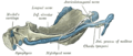 Mandible of human embryo 95 mm long. Inner aspect. Nuclei of cartilage stippled.
 -
 Mandibular division of trifacial nerve, seen from the middle line.
 -
 Inferior alveolar nerve
 -
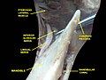 Mandibular nerve and bone. Deep dissection. Anterior view.
 -
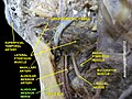
 Infratemporal fossa. Lingual and inferior alveolar nerve. Deep dissection. Anterolateral view


References
- ^ S2CID 238474299.
- ^ ISBN 978-0-8089-2371-8.
- PMID 27269666.
- ^ Fehrenbach MJ, Herring SW (2011). Illustrated Anatomy of the Head and Neck (4th ed.). Philadelphia, PA: Saunders. p. 59.
- PMID 22247929.
- PMID 24171179.
- ^ "Recovering from Surgery". Royal College of Surgeons.
- PMID 24012054.
- S2CID 21579439.
- S2CID 21328385.
- ^ "Nerve Damage Associated with Peripheral Nerve Block" (PDF). Information for patients. The Royal College of Anaesthetists. 2013. Archived from the original (PDF) on 2014-04-16. Retrieved 2014-04-15.
- ^ Tidy G (27 February 2018). Bonsall A (ed.). "Anaesthesia (UK) - Local and General anaesthesia information". Patient. Egton Medical Information Systems Limited.
- S2CID 231910002.
- PMID 27933616.
External links
- Anatomy figure: 27:03-06 at Human Anatomy Online, SUNY Downstate Medical Center
- MedEd at Loyola GrossAnatomy/h_n/cn/cn1/cnb3.htm
- lesson4 at The Anatomy Lesson by Wesley Norman (Georgetown University) (mandibularnerve)
- cranialnerves at The Anatomy Lesson by Wesley Norman (Georgetown University) (V)