
Mandibular foramen
This article includes a improve this article by introducing more precise citations. (May 2015) ) |
| Mandibular foramen | |
|---|---|
skeletal | |
| Identifiers | |
| Latin | foramen mandibulae |
| TA98 | A02.1.15.028 |
| TA2 | 865 |
| FMA | 53172 |
| Anatomical terms of bone] | |
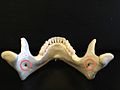
The mandibular foramen is an opening on the internal surface of the
Structure
The mandibular foramen is an opening on the internal surface of the ramus of the mandible.[1][2] It allows for divisions of the mandibular nerve and blood vessels to pass through.[2]
Variation
There are two distinct anatomies to its rim.
- In the common form the rim is V-shaped, with a groove separating the anterior and posterior parts.
- In the horizontal-oval form there is no groove, and the rim is horizontally oriented and oval in shape, the anterior and posterior parts connected.
Rarely, a bifid inferior alveolar nerve may be present, in which case a second mandibular foramen, more inferiorly placed, exists and can be detected by noting a doubled mandibular canal on a radiograph.[3]
Function
The
Clinical significance
Local anaesthetic can be injected around the mandibular foramen to anaesthetise the mandibular nerve, and thereby the mandible the lower teeth on that side, and some skin on the lower face.[1]
Other animals
The mandibular foramen can be found in other mammals, such as horses.[2]
Additional images
-
 View from behind of the mandibular foramina (red).

References
- ^ ISBN 9780323089357.
- ^ ISBN 978-0-7020-2980-6.
- ISBN 9781455706303.
External links
- cranialnerves at The Anatomy Lesson by Wesley Norman (Georgetown University) (V)
- Nicholson, Michael L. (1985). "A study of the position of the mandibular foramen in the adult human mandible". The Anatomical Record. 212 (1): 110–2. S2CID 11785956.
- Hetson, George; Share, Jack; Frommer, Jack; Kronman, Joseph H. (1988). "Statistical evaluation of the position of the mandibular foramen". Oral Surgery, Oral Medicine, Oral Pathology. 65 (1): 32–4. PMID 3422395.
- Smith, Fred H. (1978). "Evolutionary significance of the mandibular foramen area in neandertals". American Journal of Physical Anthropology. 48 (4): 523–31. PMID 96699.