
Middle cranial fossa
| Middle cranial fossa | |
|---|---|
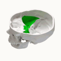
 Superior view of the skull base. Middle cranial fossa shown in green. 1: Sphenoidal limbus (anterior margin of the chiasmatic groove) lesser wings of the sphenoid 3: Dorsum sellae of the sphenoid bone 4: Superior borders of the petrous part of the temporal bone | |
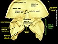
 Base of the skull. Upper surface. (Middle cranial fossa is the centermost of the three indentations, in pink and yellow.) | |
| Details | |
| Identifiers | |
| Latin | fossa cranii media |
| MeSH | D035301 |
| TA98 | A02.1.00.049 |
| TA2 | 452 |
| FMA | 54369 |
| Anatomical terminology | |
The middle cranial fossa is formed by the
It is bounded in front by the posterior margins of the lesser wings of the
Anatomy
Features

Middle part
The middle part of the fossa presents, in front, the
Behind the optic foramen the anterior clinoid process is directed backward and medialward and gives attachment to the cerebellar tentorium .
Behind the tuberculum sellae is a deep depression, the
The sella turcica is bounded posteriorly by a quadrilateral plate of bone, the
On either side of the sella turcica is the carotid groove, which is broad, shallow, and curved somewhat like the italic letter f.
It begins behind at the foramen lacerum, and ends on the medial side of the anterior clinoid process, where it is sometimes converted into a foramen (carotico-clinoid) by the union of the anterior with the middle clinoid process; posteriorly, it is bounded laterally by the lingula.
This groove lodges the
Lateral parts
The lateral parts of the middle fossa are of considerable depth, and support the
They are marked by depressions for the brain convolutions and traversed by furrows for the anterior and posterior branches of the
These furrows begin near the
The following apertures are also to be seen.
In front is the
It transmits to the
Behind the medial end of the superior orbital fissure is the foramen rotundum, for the passage of the maxillary nerve.
Behind and lateral to the foramen rotundum is the
Medial to the foramen ovale is the
Lateral to the foramen ovale is the foramen spinosum, for the passage of the middle meningeal vessels, and a recurrent branch from the mandibular nerve.
Medial to the foramen ovale is the foramen lacerum; in the fresh state the lower part of this aperture is filled up by a layer of fibrocartilage, while its upper and inner parts transmit the internal carotid artery surrounded by a plexus of sympathetic nerves.
The nerve of the pterygoid canal and a meningeal branch from the ascending pharyngeal artery pierce the layer of fibrocartilage.
On the anterior surface of the petrous portion of the temporal bone are seen the eminence caused by the projection of the
Clinical significance
A middle fossa craniotomy is one means to surgically remove acoustic neuromas (vestibular schwannoma) growing within the internal auditory canal of the temporal bone.
See also
References
![]() This article incorporates text in the public domain from page 190 of the 20th edition of Gray's Anatomy (1918)
This article incorporates text in the public domain from page 190 of the 20th edition of Gray's Anatomy (1918)
- OCLC 1201341621.)
{{cite book}}: CS1 maint: location missing publisher (link - ISBN 9781437735802.
Additional images
-
 Animation
 -
 Middle cranial fossa at human foetus
 -

-
 Middle cranial fossa
 -
 Middle cranial fossa
 -
 Middle cranial fossa


External links
- Anatomy photo:22:os-0802 at the SUNY Downstate Medical Center