
Saphenous nerve
| Saphenous nerve | |
|---|---|
 Nerves of the right lower extremity. Front view. (Saphenous labeled at center right.) | |
| Details | |
| From | femoral nerve (L3, L4) |
| Identifiers | |
| Latin | nervus saphenus |
| TA98 | A14.2.07.023 |
| TA2 | 6525 |
| FMA | 45262 |
| Anatomical terms of neuroanatomy | |
The saphenous nerve (long or internal saphenous nerve) is the largest
Structure
It is purely a sensory nerve.[2]
Origin
The saphenous nerve is the largest and terminal branch of the femoral nerve.[3] It is derived from the lumbar plexus (L3-L4).[1]
Course
Shortly after the femoral nerve passes under the
The nerve then passes along the tibial side of the leg, accompanied by the great saphenous vein.[4] It descends behind the medial border of the tibia, and, at the lower third of the leg, divides into two branches:
- one continues its course along the margin of the tibia, and ends at the ankle.
- the other passes in front of the ankle, and is distributed to the skin on the medial side of the superficial peroneal nerve.
Branches
The saphenous nerve, about the middle of the thigh, gives off a branch which joins the subsartorial plexus.
At the medial side of the knee it gives off a large
Below the knee, the branches of the saphenous nerve (medial crural cutaneous branches) are distributed to the skin of the front and medial side of the leg, communicating with the cutaneous branches of the femoral, or with filaments from the obturator nerve.
Clinical significance
Procedures such as
The saphenous nerve can experience
Additional images
-
 Cross-section through the middle of the thigh.
 -
 Cross-section through middle of leg.
 -
 The femoral artery.
 -
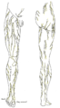 Cutaneous nerves of the right lower extremity. Front and posterior views.
 -
 Cutaneous nerves of the right lower extremity. Front and posterior views.
 -
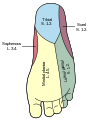 Diagram of the segmental distribution of the cutaneous nerves of the sole of the foot.
 -
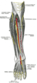 Deep nerves of the front of the leg.
 -
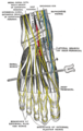 Nerves of the dorsum of the foot.


References
![]() This article incorporates text in the public domain from page 956 of the 20th edition of Gray's Anatomy (1918)
This article incorporates text in the public domain from page 956 of the 20th edition of Gray's Anatomy (1918)
- ^ PMID 31082089, retrieved 11 January 2023
- ^ ISBN 978-0-443-06651-1, retrieved 21 February 2021
- ^ ISBN 978-0-12-226870-0, retrieved 21 February 2021
- ^ ISBN 978-0-12-226870-0, retrieved 21 February 2021
- PMID 22096447.
- ^ Brad McKechnie (22 May 1995). "Saphenous Nerve Entrapment Neuropathy". Dynamic Chiropractic. 13 (11).
External links
- Anatomy photo:12:08-0102 at the SUNY Downstate Medical Center - "Structures of the Adductor Canal"