
Medial plantar nerve
| Medial plantar nerve | |
|---|---|
 The plantar nerves. | |
| Details | |
| From | Tibial nerve |
| Innervates | Sole, Abductor hallucis muscle, Flexor digitorum brevis muscle, Flexor hallucis brevis muscle, 1 medial lumbrical |
| Identifiers | |
| Latin | nervus plantaris medialis |
| TA98 | A14.2.07.066 |
| TA2 | 6590 |
| FMA | 44716 |
| Anatomical terms of neuroanatomy | |
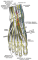
The medial plantar nerve (internal plantar nerve) is the larger of the two terminal divisions of the tibial nerve (medial and lateral plantar nerve), which accompanies the medial plantar artery.
From its origin under the
Branches
The branches of the medial plantar nerve are: (1) cutaneous, (2) muscular, (3) articular, (4) a proper digital nerve to the medial side of the great toe, and (5) three common digital nerves.
Cutaneous branches
The cutaneous branches pierce the
Muscular branches
The muscular branches supply muscles on the medial side of the sole, including the abductor hallucis, the flexor digitorum brevis, the flexor hallucis brevis, and the first
Articular branches
The articular branches supply the
Proper digital nerve of the great toe
The proper digital nerve of the
Three common digital nerves
The three common digital nerves (nn. digitales plantares communes) pass between the divisions of the plantar aponeurosis, and each splits into two proper digital nerves—those of the first common digital nerve supply the adjacent sides of the great and second toes; those of the second, the adjacent sides of the second and third toes; and those of the third, the adjacent sides of the third and fourth toes.
The third common digital nerve receives a communicating branch from the lateral plantar nerve; the first gives a twig to the first lumbricals.
Each proper digital nerve gives off cutaneous and articular filaments; and opposite the last phalanx sends upward a dorsal branch, which supplies the structures around the nail, the continuation of the nerve being distributed to the ball of the toe.
It will be observed that these digital nerves are similar in their distribution to those of the median nerve in the hand.
Additional images
-
 Coronal section through right talocrural and talocalcaneal joints.
 -
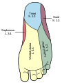 Diagram of the segmental distribution of the cutaneous nerves of the sole of the foot.
 -
 Nerves of the dorsum of the foot.


References
![]() This article incorporates text in the public domain from page 963 of the 20th edition of Gray's Anatomy (1918)
This article incorporates text in the public domain from page 963 of the 20th edition of Gray's Anatomy (1918)