
Perineal nerve
| Perineal nerve | |
|---|---|
superficial transverse perineal muscle, bulbospongiosus muscle, ischiocavernosus muscle, bulb of penis, levator ani, external anal sphincter | |
| Identifiers | |
| Latin | nervi perineales |
| TA98 | A14.2.07.039 |
| TA2 | 6556 |
| FMA | 21866 |
| Anatomical terms of neuroanatomy] | |
The perineal nerve is a
deep branch to muscles. It supplies the skin and muscles of the perineum. Its latency is tested with electrodes
.
Structure
The perineal nerve is a branch of the
deep branch of the perineal nerve (also known as the "muscular" branch) travels to the muscles of the perineum.[1] Both of these are superficial to the dorsal nerve of the penis or the dorsal nerve of the clitoris.[4]
Function
The perineal nerve supplies the skin and muscles of the
superficial transverse perineal muscle, the bulbospongiosus muscle, the ischiocavernosus muscle, the bulb of penis, levator ani, and the external anal sphincter.[1]
Clinical significance
The latency of the perineal nerve can be measured with electrodes.[5] It is used to test nerve function.[5]
Additional images
-
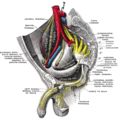 Sacral plexus of the right side. (Perineal nerve visible at center right.)

References
![]() This article incorporates text in the public domain from page 968 of the 20th edition of Gray's Anatomy (1918)
This article incorporates text in the public domain from page 968 of the 20th edition of Gray's Anatomy (1918)
- ^ ISBN 978-0-12-803062-2.
- ^ ISBN 978-1-4160-4572-4.
- ^ Essential Clinical Anatomy. K.L. Moore & A.M. Agur. Lippincott, 2 ed. 2002. Page 263
- PMID 26003239.
- ^ ISBN 978-0-443-06707-5.
External links
- Anatomy photo:41:10-0100 at the SUNY Downstate Medical Center - "The Female Perineum: The Perineal Nerve"
- Anatomy image:9174 at the SUNY Downstate Medical Center
- Anatomy image:9187 at the SUNY Downstate Medical Center
- figures/chapter_32/32-3.HTM: Basic Human Anatomy at Dartmouth Medical School