
Spiral ganglion
| Spiral ganglion | |
|---|---|
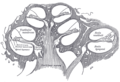
 Transverse section of the cochlear duct of a fetal cat. (Ganglion spirale is labeled at top, second from left.) | |
 Part of the cochlear division of the acoustic nerve, highly magnified. | |
| Details | |
| Identifiers | |
| Latin | ganglion spirale |
| MeSH | D013136 |
| TA98 | A15.3.03.125 |
| TA2 | 6319 |
| TH | H3.11.09.3.04068 |
| FMA | 53445 |
| Anatomical terminology | |
The spiral (cochlear) ganglion is a group of neuron cell bodies in the modiolus, the conical central axis of the cochlea. These bipolar neurons innervate the hair cells of the organ of Corti. They project their axons to the ventral and dorsal cochlear nuclei as the cochlear nerve, a branch of the vestibulocochlear nerve (CN VIII).
Structure
eighth cranial nerve. The number of neurons in the spiral ganglion is estimated to be about 35,000–50,000.[1]
Two apparent subtypes of spiral ganglion cells exist. Type I spiral ganglion cells comprise the vast majority of spiral ganglion cells (90-95% in cats and 88% in humans
Development
The
auditory vesicle. The ganglion gradually splits into two parts, the vestibular ganglion and the spiral ganglion. The axons of neurons in the spiral ganglion travel to the brainstem, forming the cochlear nerve
.
Gallery
-
 Diagrammatic longitudinal section of the cochlea
 -
 Organ of corti


References
![]() This article incorporates text in the public domain from page 1051 of the 20th edition of Gray's Anatomy (1918)
This article incorporates text in the public domain from page 1051 of the 20th edition of Gray's Anatomy (1918)
- ISBN 0-7817-6003-8.
- ISBN 0-387-97800-3.
- PMID 5015157.
- ^ JB Nadol Jr (1990). "Synaptic morphology of inner and outer hair cells of the human organ of Corti". J Elect Micr Tech.
External links
- Slide and overview at anatomy.dal.ca
- Slide at cytochemistry.net
- Image at University of New England, Maine