
Organ of Corti
This article needs additional citations for verification. (December 2014) |
| Organ of Corti | |
|---|---|
 A cross section of the cochlea illustrating the organ of Corti | |
| Details | |
| Part of | Cochlea of the inner ear |
| Identifiers | |
| Latin | organum spirale |
| MeSH | D009925 |
| NeuroLex ID | birnlex_2526 |
| TA98 | A15.3.03.121 |
| TA2 | 7035 |
| FMA | 75715 |
| Anatomical terminology | |
The organ of Corti, or spiral organ, is the receptor organ for hearing and is located in the mammalian cochlea. This highly varied strip of epithelial cells allows for transduction of auditory signals into nerve impulses' action potential.[1] Transduction occurs through vibrations of structures in the inner ear causing displacement of cochlear fluid and movement of hair cells at the organ of Corti to produce electrochemical signals.[2]
Italian anatomist Alfonso Giacomo Gaspare Corti (1822–1876) discovered the organ of Corti in 1851.[3] The structure evolved from the basilar papilla and is crucial for mechanotransduction in mammals.
Structure

The organ of Corti is located in the
Projecting from the tops of the hair cells are tiny finger-like projections called stereocilia, which are arranged in a graduated fashion with the shortest stereocilia on the outer rows and the longest in the center. This gradation is thought to be the most important anatomic feature of the organ of Corti because this allows the sensory cells superior tuning capability.[5]
If the cochlea were uncoiled, it would roll out to be about 33 mm long in women and 34 mm in men, with about 2.28 mm of standard deviation for the population.[6] The cochlea is also tonotopically organized, meaning that different frequencies of sound waves interact with different locations on the structure. The base of the cochlea, closest to the outer ear, is the most stiff and narrow and is where the high-frequency sounds are transduced. The apex, or top, of the cochlea is wider and much more flexible and loose and functions as the transduction site for low-frequency sounds.[7]
Function

The function of the organ of Corti is to convert (transduce) sounds into electrical signals that can be transmitted to the brainstem through the auditory nerve.[2] It is the auricle and middle ear that act as mechanical transformers and amplifiers so that the sound waves end up with amplitudes 22 times greater than when they entered the ear.
Auditory transduction
In normal hearing, the majority of the auditory signals that reach the organ of Corti in the first place come from the outer ear.
The basilar membrane on the tympanic duct presses against the hair cells of the organ as
Cochlear amplification
The organ of Corti is also capable of modulating the auditory signal.[7] The outer hair cells (OHCs) can amplify the signal through a process called electromotility where they increase movement of the basilar and tectorial membranes and therefore increase deflection of stereocilia in the IHCs.[8][10][11]
A crucial piece to this cochlear amplification is the motor protein prestin, which changes shape based on the voltage potential inside of the hair cell. When the cell is depolarized, prestin shortens, and because it is located on the membrane of OHCs it then pulls on the basilar membrane and increasing how much the membrane is deflected, creating a more intense effect on the inner hair cells (IHCs). When the cell hyperpolarizes prestin lengthens and eases tension on the IHCs, which decreases the neural impulses to the brain. In this way, the hair cell itself is able to modify the auditory signal before it even reaches the brain.
Development
The organ of Corti, in between the
Development and growth of the organ of Corti relies on specific genes, many of which have been identified in previous research (
to undergo such differentiation. Specifically, the cochlear duct growth and the formation of hair cells within the organ of Corti.Mutations in the genes expressed in or near the organ of Corti before the differentiation of hair cells will result in a disruption in the differentiation, and potential malfunction of, the organ of Corti.
Clinical significance
Hearing loss
The organ of Corti can be damaged by excessive sound levels, leading to
The most common kind of hearing impairment,
While hearing loss has always been considered irreversible in mammals, fish and birds routinely repair such damage. A 2013 study has shown that the use of particular drugs may reactivate genes normally expressed only during hair cell development. The research was carried out at Harvard Medical School, Massachusetts Eye and Ear, and the Keio University School of Medicine in Japan.[14][15]
Additional images
-
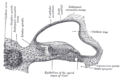 Transverse section of the cochlear duct of a fetal cat.
 -
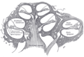 Diagrammatic longitudinal section of the cochlea
 -
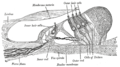
 Floor of ductus cochlearis
 -
 Limbus laminæ spiralis and membrana basilaris
 -
 Section through the spiral organ of Corti (magnified)


Notes
- S2CID 3716179.
- ^ a b c The Ear Pujol, R., Irving, S., 2013
- PMID 18652163.
- ^ S2CID 2962483.
- S2CID 4764624.
- PMID 17471760.
- ^ PMID 21256948.
- ^ ISBN 978-0-87893-609-0.
- S2CID 3506274.
- PMID 3656195.

- S2CID 17722638.
- PMID 3515985.
- ISBN 0-7693-0052-9.
- ^ "Cochlear hair cells - Beyond the Dish". wordpress.com.
- PMID 26157030.
References
- Corti A (1851). "Recherches sur l'organe de l'ouïe des mammiferes". Zeitschrift für Wissenschaftliche Zoologie. 3: 106–169.
- Fritzsch B, Jahan I, Pan N, Kersigo J, Duncan J, Kopecky B (2012). "Dissecting the molecular basis of organ of Corti development: where are we now?". Hearing Research. 276 (1–2): 16–26. PMID 21256948.
History. (n.d.).
- Hudspeth A (2014). "Integrating the active process of hair cells with cochlear function". Nature Reviews Neuroscience. 15 (9): 600–614. S2CID 3716179.
- Lim D (1986). "Functional structure of the organ of Corti: a review". Hearing Research. 22 (1–3): 117–146. S2CID 4764624.
- Malgrange B, Thiry M, Van de Water TR, Nguyen L, Moonen G, Lefebvre PP (2002). "Epithelial supporting cells can differentiate into outer hair cells and Deiters' cells in the cultured organ of Corti". Cellular and Molecular Life Sciences. 59 (10): 1744–1757. S2CID 2962483.
- Nicholls, J. G., Martin, A. R., Fuchs, P. A., Brown, D. A., Diamond, M. E., & Weisblat, D. A. (2012). From Neuron to Brain (5th ed., pp. 456–459). Sunderland, MA: Sinauer Associates, Inc.
- Pritchard U. "On the organ of Corti in mammals". 2 March 1876, OCLC 1778190
- Pujol, R., & Irving, S. (2013). The Ear.
External links
- Dissecting the molecular basis of organ of Corti development PMC 3097286
- Organ of Corti 3D animation
- http://lobe.ibme.utoronto.ca/presentations/OHC_Electromotility/sld005.htm Archived 2005-03-30 at the Wayback Machine Diagram at University of Toronto
- http://mayoresearch.mayo.edu/mayo/research/ent_research/images/image02.gif Archived 2007-03-05 at the Wayback Machine Diagram at Mayo
- http://www.iurc.montp.inserm.fr/cric51/audition/english/corti/fcorti.htm Archived 2006-11-30 at the Wayback Machine at University of Montpellier 1
See also
| National | |
|---|---|
| Other | |