Vorticose veins
| Vorticose veins | |
|---|---|
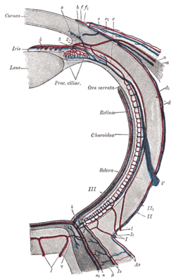
 The veins of the choroid. (Venae vorticosae labeled - though difficult to see - at center.) | |
 Diagram of the blood vessels of the eye, as seen in a horizontal section. ("V", at center right, is the label for the vena vorticosa) | |
| Details | |
| Drains to | Superior ophthalmic vein, and inferior ophthalmic vein |
| Artery | short posterior ciliary arteries[citation needed] |
| Identifiers | |
| Latin | venae vorticosae |
| TA98 | A12.3.06.106 |
| TA2 | 4892 |
| FMA | 70880 |
| Anatomical terminology | |
The vorticose veins, referred to clinically as the vortex veins,[1] are veins that drain the choroid of the eye. There are usually 4-5 vorticose veins in each eye, with at least one vorticose vein per each quadrant of the eye. Vorticose veins drain into the superior ophthalmic vein, and inferior ophthalmic vein.[2]
Vorticose veins are an important ophthalmoscopic landmark.[3]
Structure
Course and relations
Vorticose veins exit the eyeball 6 mm posterior to its equator.[2]
Fate
Upper vortex veins empty into the superior ophthalmic vein, and lower vortex veins empty into the inferior ophthalmic vein.[2][4]
Variation
The number of vorticose veins is known to vary from 4 to 8, with about 65% of the normal population having 4 or 5[1] with at least one vein in each quadrant.[2]
Clinical significance
Vorticose veins are an important ophthalmoscopic landmark.[3] They can be visualised in a dilated pupil using an indirect ophthalmoscope.[2]
Additional images
-
The blood-vessels of the eyeball (diagrammatic).
References
- ^ S2CID 42756249.
- ^ ISBN 978-1-4377-1926-0.
- ^ PMID 6512144.
- OCLC 1201341621.)
{{cite book}}: CS1 maint: location missing publisher (link
External links

