
Brachial plexus
| Brachial plexus | |
|---|---|
 The right brachial plexus with its short branches, viewed from in front. | |
 The roots, trunks and cords of the plexus shown in a dissected cadavaric specimen. | |
| Details | |
| Function | Network (nerve plexus) of nerves that supply the arms. |
| Identifiers | |
| Latin | plexus brachialis |
| MeSH | D001917 |
| TA98 | A14.2.03.001 |
| TA2 | 6395 |
| FMA | 5906 |
| Anatomical terms of neuroanatomy | |
The brachial plexus is a network of nerves (
Structure
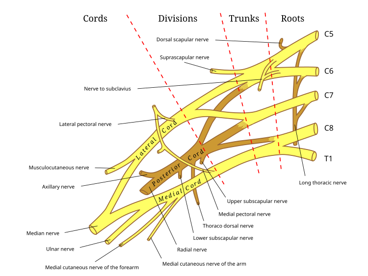
The brachial plexus is divided into five roots, three trunks, six divisions (three anterior and three posterior), three cords, and five branches. There are five "terminal" branches and numerous other "pre-terminal" or "collateral" branches, such as the subscapular nerve, the thoracodorsal nerve, and the long thoracic nerve,[1] that leave the plexus at various points along its length.[2] A common structure used to identify part of the brachial plexus in cadaver dissections is the M or W shape made by the musculocutaneous nerve, lateral cord, median nerve, medial cord, and ulnar nerve.
Roots
The five roots are the five
Trunks
These roots merge to form the trunks:
Divisions
Each trunk then splits in two, to form six divisions:
- anterior divisions of the upper, middle, and lower trunks
- posterior divisions of the upper, middle, and lower trunks
- when observing the body in the anatomical position, the anterior divisions are superficial to the posterior divisions
Cords
These six divisions regroup to become the three cords or large fiber bundles. The cords are named by their position with respect to the axillary artery.
- The posterior cord is formed from the three posterior divisions of the trunks (C5-C8, T1)
- The lateral cord is formed from the anterior divisions of the upper and middle trunks (C5-C7)
- The medial cord is simply a continuation of the anterior division of the lower trunk (C8, T1)
Diagram
Branches
The branches are listed below. Most branches arise from the cords, but few branches arise (indicated in italics) directly from earlier structures. The five on the left are considered "terminal branches". These terminal branches are the musculocutaneous nerve, the axillary nerve, the radial nerve, the median nerve, and the ulnar nerve. Due to both emerging from the lateral cord the musculocutaneous nerve and the median nerve are well connected. The musculocutaneous nerve has even been shown to send a branch to the median nerve further connecting them.[1] There have been several variations reported in the branching pattern but these are very rare.[3]
Bold indicates primary spinal root component of nerve. Italics indicate spinal roots that frequently, but not always, contribute to the nerve.
| From | Nerve | Roots[4] | Muscles | Cutaneous |
| roots | dorsal scapular nerve | C4, C5 | levator scapulae |
- |
| roots | long thoracic nerve | C5, C6, C7 | serratus anterior |
- |
| roots | branch to phrenic nerve | C3, C4,C5 | Diaphragm | - |
| upper trunk | nerve to the subclavius |
C5, C6 | subclavius muscle | - |
| upper trunk | suprascapular nerve | C5, C6 | infraspinatus |
- |
| lateral cord | lateral pectoral nerve | C5, C6, C7 | pectoralis major and pectoralis minor (by communicating with the medial pectoral nerve) | - |
| lateral cord | musculocutaneous nerve | C5, C6, C7 | biceps brachii |
Becomes the lateral cutaneous nerve of the forearm Innervates the skin of the anterolateral forearm; elbow joint.[2]
|
| lateral cord | lateral root of the median nerve | C5,C6,C7 | fibres to the median nerve
(see below) |
- |
| posterior cord | upper subscapular nerve | C5, C6 | subscapularis (upper part) |
- |
| posterior cord | thoracodorsal nerve (middle subscapular nerve) | C6, C7, C8 | latissimus dorsi |
- |
| posterior cord | lower subscapular nerve | C5, C6 | subscapularis (lower part ) and teres major |
- |
| posterior cord | axillary nerve | C5, C6 | anterior branch: teres minor and deltoid muscles |
posterior branch becomes superior lateral cutaneous nerve of arm Innervates the skin of the lateral shoulder and arm: shoulder joint.[2] |
| posterior cord | radial nerve | C5, C6, C7, C8, T1 | extensor muscles of the forearm, and brachioradialis |
skin of the posterior arm and posterior forearm as the posterior cutaneous nerve of the arm and the posterior cutaneous nerve of the forearm, respectively. Also superficial branch of radial nerve supplies back of the hand, including the web of skin between the thumb and index finger. |
| medial cord | medial pectoral nerve | C8, T1 | pectoralis major and pectoralis minor | - |
| medial cord | medial root of the median nerve | C8, T1 | all of the flexor digitorum profundus that supplies the 2nd and 3rd digits
1st and 2nd lumbrical muscles. muscles of the thenar eminence by a recurrent thenar branch |
portions of hand not served by ulnar or radial, i.e skin of the palmar side of the thumb, the index and middle finger, half the ring finger, and the nail bed of these fingers |
| medial cord | medial cutaneous nerve of the arm |
C8, T1 | - | front and medial skin of the arm |
| medial cord | medial cutaneous nerve of the forearm |
C8, T1 | - | medial skin of the forearm |
| medial cord | ulnar nerve | C7, C8, T1(C7 because it supplies to the Flexor carpi ulnaris) | thenar muscles and the two lateral lumbricals of the hand which are served by the median nerve |
the skin of the medial side of the hand and medial one and a half fingers on the palmar side and medial two and a half fingers on the dorsal side |
Function
The brachial plexus provides nerve supply to the skin and muscles of the arms, with two exceptions: the
The terminal branches of the brachial plexus (musculocutaneous n., axillary n., radial n., median n., and ulnar n.) all have specific sensory, motor and proprioceptive functions.[5][6]
| Terminal Branch | Sensory Innervation | Muscular Innervation |
|---|---|---|
| musculocutaneous nerve | Skin of the anterolateral forearm | Brachialis, biceps brachii, coracobrachialis |
| axillary nerve | Skin of lateral portion of the shoulder and upper arm | Deltoid and teres minor |
| radial nerve | Posterior aspect of the lateral forearm and wrist; posterior arm | Triceps brachii, brachioradialis, anconeus, extensor muscles of the posterior arm and forearm |
| median nerve | Skin of lateral 2/3rd of hand and the tips of digits 1-4 | Forearm flexors, thenar eminence, lumbricals of the hand 1-2 |
| ulnar nerve | Skin of palm and medial side of hand and digits 3-5 | Hypothenar eminence, some forearm flexors, thumb adductor, lumbricals 3-4, interosseous muscles |
Clinical significance
Injury


Injury to the brachial plexus may affect sensation or movement of different parts of the arm. Injury can be caused by the shoulder being pushed down and the head being pulled up, which stretches or tears the nerves. Injuries associated with malpositioning commonly affect the brachial plexus nerves, rather than other peripheral nerve groups.[7][8] Due to the brachial plexus nerves being very sensitive to position, there are very limited ways of preventing such injuries. The most common victims of brachial plexus injuries consist of victims of motor vehicle accidents and newborns.[9]
Injuries can be caused by stretching, diseases, and wounds to the lateral cervical region (posterior triangle) of the neck or the axilla. Depending on the location of the injury, the signs and symptoms can range from complete paralysis to anesthesia. Testing the patient's ability to perform movements and comparing it to their normal side is a method to assess the degree of paralysis. A common brachial plexus injury is from a hard landing where the shoulder widely separates from the neck (such as in the case of motorcycle accidents or falling from a tree). These stretches can cause ruptures to the superior portions of the brachial plexus or avulse the roots from the spinal cord. Upper brachial plexus injuries are frequent in newborns when excessive stretching of the neck occurs during delivery. Studies have shown a relationship between a newborn's weight and brachial plexus injuries; however, the number of cesarean deliveries necessary to prevent a single injury is high at most birth weights.[10]
For the upper brachial plexus injuries, paralysis occurs in those muscles supplied by C5 and C6 like the deltoid, biceps, brachialis, and brachioradialis. A loss of sensation in the lateral aspect of the upper limb is also common with such injuries. An inferior brachial plexus injury is far less common but can occur when a person grasps something to break a fall or a baby's upper limb is pulled excessively during delivery. In this case, the short muscles of the hand would be affected and cause the inability to form a full fist position.[11]
To differentiate between preganglionic and postganglionic injury, clinical examination requires that the physician keep the following points in mind. Preganglionic injuries cause loss of sensation above the level of the clavicle, pain in an otherwise insensate hand, ipsilateral Horner's syndrome, and loss of function of muscles supplied by branches arising directly from roots—i.e., long thoracic nerve palsy leading to winging of scapula and elevation of ipsilateral diaphragm due to phrenic nerve palsy.
Acute brachial plexus neuritis is a neurological disorder that is characterized by the onset of severe pain in the shoulder region. Additionally, the compression of cords can cause pain radiating down the arm, numbness, paresthesia, erythema, and weakness of the hands. This kind of injury is common for people who have prolonged hyperabduction of the arm when they are performing tasks above their head.
Sports injuries
One sports injury that is becoming prevalent in contact sports, particularly in the sport of American football, is called a "stinger." An athlete can incur this injury in a collision that can cause cervical axial compression, flexion, or extension of nerve roots or terminal branches of the brachial plexus.
Penetrating wounds
Most penetration wounds require immediate treatment and are not as easy to repair. For example, a deep knife wound to the brachial plexus could damage and/or sever the nerve. According to where the cut was made, it could inhibit action potentials needed to innervate that nerve's specific muscle or muscles.
Injuries during birth
Brachial plexus injuries can occur during the delivery of newborns when after the delivery of the head, the anterior shoulder of the infant cannot pass below the pubic symphysis without manipulation. This manipulation can cause the baby's shoulder to stretch, which can damage the brachial plexus to varying degrees.
Tumors
Tumors that may occur in the brachial plexus are schwannomas, neurofibromas and malignant peripheral nerve sheath tumors.
Imaging
Imaging of the Brachial Plexus can be done effectively by using a higher magnetic strength MRI Scanner like 1.5 T or more. It is impossible to evaluate the brachial plexuses with plain Xray, CT and ultrasound scanning can manage to view the plexuses to an extent; hence MRI is preferred in imaging brachial plexus over other imaging modalities due to its multiplanar capability and the tissue contrast difference between brachial plexus and adjacent vessels. The plexuses are best imaged in coronal and sagittal planes, but axial images give an idea about the nerve roots. Generally, T1 WI and T2 WI images are used in various planes for the imaging; but new sequences like MR Myelolography, Fiesta 3D and T2 cube are also used in addition to the basic sequences to gather more information to evaluate the anatomy more.
In anaesthetics
See also
- Plexus
- Nerve plexus
- Cranial nerve
- Spinal nerve
- List of anatomy mnemonics
Additional images
-
 The brachial plexus surrounds the brachial artery.
 -
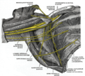 Nerves in the infraclavicular portion of the right brachial plexus in the axillary fossa.
 -
 The outermost (distal) part of the brachial plexus shown from a dissected cadaveric specimen.
 -
 Brachial plexus
 -
 Mind map showing branches of brachial plexus
 -
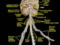 Spinal cord. Brachial plexus. Cerebrum.Inferior view.Deep dissection.
 -
 Diagram of the brachial plexus using colour to illustrate the contributions of each nerve root to the branches.
 -
 The brachial plexus, including all branches of the C5-T1 ventral primary rami. Includes mnemonics for learning the plexus's connections and branches.
 -
 Mixed fibres of a spinal nerve


References
- ^ ISBN 9810231393.
- ^ ISBN 9789814646437.
- PMID 24693486.
- ISBN 978-0-7817-6274-8.
- ISBN 9789814646437.
- ^ "Axillary Brachial Plexus Block". www.nysora.com. New York School of Regional Anesthesia. 2013-09-20. Archived from the original on 2017-07-12.
- PMID 3342585.
- PMID 23307682.
- PMID 9179891.
- PMID 9166293.
- ISBN 0-7817-3639-0.
- PMID 17553154. Retrieved 2015-02-06.
- . Retrieved 2015-02-12.
- ^ "Brachial Plexus Injuries Information Page: National Institute of Neurological Disorders and Stroke (NINDS)". www.ninds.nih.gov. Archived from the original on 2016-12-02. Retrieved 2016-11-28.
- PMID 19691859.
Bibliography
- Saladin, Kenneth (2014). Anatomy and Physiology (7th ed.). McGraw-Hill Education. p. 491.
- Kishner, Stephen. "Brachial Plexus Anatomy". Medscape. WebMD. Retrieved 29 Nov 2015.
External links
- Brachial Plexus Injury/Illustration, Cincinnati Children's Hospital Medical Center
- Learn the Brachial Plexus in Five Minutes or Less by Daniel S. Romm, M.D. and Dennis A. Chu Chu, M.D. [1]
- Video of the dissected axilla and brachial plexus