
Lumbricals of the hand
| Lumbricals of the hand | |
|---|---|
interphalangeal joints | |
| Identifiers | |
| Latin | musculi lumbricales manus |
| TA98 | A04.6.02.065 |
| TA2 | 2532 |
| FMA | 37385 |
| Anatomical terms of muscle] | |
The lumbricals are intrinsic
The
Structure
The lumbricals are four, small, worm-like muscles on each hand. These muscles are unusual in that they do not attach to bone. Instead, they attach proximally to the tendons of
| # | Form | Origin | Insertion |
| First | unipennate |
It originates from the radial side of the most radial tendon of the flexor digitorum profundus (corresponding to the index finger). | It passes posteriorly along the radial side of the index finger to insert on the extensor expansion near the metacarpophalangeal joint. |
| Second | unipennate | It originates from the radial side of the second most radial tendon of the flexor digitorum profundus (which corresponds to the middle finger). | It passes posteriorly along the radial side of the middle finger and inserts on the extensor expansion near the metacarpophalangeal joint. |
| Third | bipennate |
One head originates on the radial side of the flexor digitorum profundus tendon corresponding to the ring finger, while the other originates on the ulnar side of the tendon for the middle finger. | The muscle passes posteriorly along the radial side of the ring finger to insert on its extensor expansion. |
| Fourth | bipennate | One head originates on the radial side of the flexor digitorum profundus tendon corresponding to the little finger, while the other originates on the ulnar side of the tendon for the ring finger. | The muscle passes posteriorly along the radial side of the little finger to insert on its extensor expansion. |

Nerve supply
The first and second lumbricals (the most radial two) are innervated by the median nerve. The third and fourth lumbricals (most ulnar two) are innervated by the deep branch of ulnar nerve.[5]
This is the usual innervation of the lumbricals (occurring in 60% of individuals). However 1:3 (median:ulnar - 20% of individuals) and 3:1 (median:ulnar - 20% of individuals) also exist. The lumbrical innervation always follows the innervation pattern of the associated muscle unit of flexor digitorum profundus (i.e. if the muscle units supplying the tendon to the middle finger are innervated by the median nerve, the second lumbrical will also be innervated by the median nerve).[6]
Blood supply
Four separate sources supply blood to these muscles: the
Function
The lumbrical muscles, with the help of the interosseous muscles, simultaneously flex the
Etymology
The term "lumbrical" comes from the Latin, meaning "worm".[8]
Additional images
-
 Tendons of forefinger and vincula tendina
 -
 Lumbricals of the hand
 -
 Lumbricals of the hand
 -
 Lumbricals muscle
 -
 Lumbricals muscle
 -
 Lumbricals muscle
 -
 Lumbricals muscle
 -
 Lumbricals muscle
 -
 Muscles of hand, cross section
 -
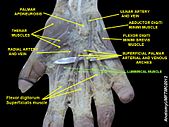
 Wrist joint. Deep dissection.Anterior, palmar view
 -
 Wrist joint. Deep dissection.Anterior, palmar view
 -
 Wrist joint. Deep dissection.Anterior, palmar view


References
- ^ ISBN 978-0-7234-3451-1. p. 97
- ^ PMID 17825776.
- ^ PMID 24369943.
- S2CID 244111.
- S2CID 8084761.
- ISBN 978-0-443-05611-6.
- S2CID 26384944.
- S2CID 81678124.