
Intercostal nerves
This article includes a list of general references, but it lacks sufficient corresponding inline citations. (February 2008) |
| Intercostal nerves | |
|---|---|
intercostal muscle | |
| Identifiers | |
| Latin | nervi intercostales |
| MeSH | D007367 |
| TA98 | A14.2.04.006 |
| TA2 | 6471 |
| FMA | 75467 |
| Anatomical terms of neuroanatomy] | |
The intercostal nerves are part of the
The first two nerves supply fibers to the
Unlike the nerves from the
The 1st thoracic nerve
The anterior division of the first thoracic nerve divides into two branches. The larger branch leaves the thorax in front of the neck of the first rib, and enters the brachial plexus. The smaller branch, the first intercostal nerve, runs along the first intercostal space, and ends on the front of the chest as the first anterior cutaneous branch of the thorax. Occasionally, this anterior cutaneous branch is missing.
The first intercostal nerve rarely gives off a lateral cutaneous branch; but sometimes sends a small branch to communicate with the intercostobrachial.
From the second thoracic nerve it frequently receives a connecting twig, which ascends over the neck of the second rib. This nerve was first described by Kuntz in 1927. There is considerable anatomic variation, but Kuntz nerve may be present in 40-80% of the population.[4][5]
The upper thoracic nerves: 2nd–6th
The anterior divisions of the second, third, fourth, fifth, and sixth thoracic nerves, and the small branch from the first thoracic, are confined to the walls of the thorax, and are named thoracic intercostal nerves.
They pass forward in the intercostal spaces below the intercostal vessels. At the back of the chest they lie between the pleura and the posterior intercostal membranes, but soon they run between the internal intercostals and the innermost intercostals then anteriorly they lie between the pleura and the internal intercostals.
Near the sternum, they cross in front of the
The fourth intercostal nerve is innervated by cutaneous slowly-adapting and rapidly-adapting mechanoreceptors, especially by ones densely-packed under the areola; innervation subsequently triggers oxytocin release, which, when in the peripheral bloodstream, causes myoepithelial cell contraction and lactation: this is an example of a non-nerve-innervation muscular reflex.
Branches
Numerous slender muscular filaments supply the
- Lateral cutaneous branches (rami cutanei laterales) are derived from the intercostal nerves, about midway between the vertebræ and sternum; they pierce the Serratus anterior, and divide into anterior and posterior branches.
- The anterior branches run forward to the side and the forepart of the chest and skin, fourth nerve anterior branches supplying the Obliquus externus abdominis.
- The posterior branches run backward, and supply the skin over the Latissimus dorsi.
The lateral cutaneous branch of the second intercostal nerve does not divide, like the others, into an anterior and a posterior branch; it is named the intercostobrachial nerve.
The lower thoracic nerves: 7th–11th
The lower thoracic nerves: 12th
Anterior division
Lateral cutaneous branch
The lateral cutaneous branch of the last thoracic nerve is large, and undivided.
It perforates the internal and the external oblique muscles, descends over the iliac crest in front of the lateral cutaneous branch of the iliohypogastric nerve, and is distributed to the skin of the front part of the gluteal muscles, some of its filaments extending as low as the greater trochanter of the femur.
Additional images
-
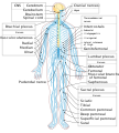 Nervous system
 -
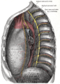 Intercostal spaces, viewed from the left
 -
 Brachial plexus
 -
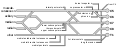
 Brachial plexus with courses of spinal nerves shown


See also
- Intercostal nerve block
- External intercostal muscles
- Internal intercostal muscles
- Peripheral nervous system
References
![]() This article incorporates text in the public domain from page 945 of the 20th edition of Gray's Anatomy (1918)
This article incorporates text in the public domain from page 945 of the 20th edition of Gray's Anatomy (1918)
- ^ ISBN 978-0-12-802653-3, retrieved 2020-11-17
- ^ ISBN 978-1-4377-0575-1, retrieved 2020-11-17
- ^ ISBN 978-0-323-08340-9, retrieved 2020-11-17
- ^ Ramsaroop L, Partab P, Singh B, Satyapal KS. Thoracic origin of a sympathetic supply to the upper limb: the 'nerve of Kuntz' revisited. J Anat. 2001;199:675Y682
- ^ Marhold F, Izay B, Zacherl J, Tschabitscher M, Neumayer C. Thoracoscopic and anatomic landmarks of Kuntz's nerve: implications for sympathetic surgery. Ann Thorac Surg. 2008;86:1653Y1658.
External links
- thoraxlesson5 at The Anatomy Lesson by Wesley Norman (Georgetown University) (paravertebralregion)
- Atlas image: abdo_wall53 at the University of Michigan Health System - "Abdominal Wall, Dissection, Lateral View"