
Lateral meniscus
This article includes a list of general references, but it lacks sufficient corresponding inline citations. (June 2015) |
| Lateral meniscus | |
|---|---|
 Knee from the side, with lateral meniscus simply labeled as "meniscus". | |
 | |
| Details | |
| Identifiers | |
| Latin | meniscus lateralis |
| TA98 | A03.6.08.002 |
| TA2 | 1885 |
| FMA | 44631 |
| Anatomical terminology | |
The lateral meniscus (external semilunar fibrocartilage) is a
Structure
The lateral meniscus is grooved laterally for the tendon of the
Its anterior end is attached in front of the intercondyloid eminence of the tibia, lateral to, and behind, the anterior cruciate ligament, with which it blends; the posterior end is attached behind the intercondyloid eminence of the tibia and in front of the posterior end of the medial meniscus.
The anterior attachment of the lateral meniscus is twisted on itself so that its free margin looks backward and upward, its anterior end resting on a sloping shelf of bone on the front of the lateral process of the
Close to its posterior attachment it sends off a strong
The lateral meniscus gives off from its anterior convex margin a fasciculus which forms the transverse ligament.
Variation
Occasionally a small fasciculus passes forward to be inserted into the lateral part of the anterior cruciate ligament.
Clinical significance
The lateral meniscus is less likely to be injured or torn than the medial meniscus. Diagnosis of lateral meniscus tear is done with
Additional images
This gallery of anatomic features needs cleanup to abide by the medical manual of style. ; please improve or remove the gallery accordingly. (June 2015) |
-
 Left knee joint from behind, showing interior ligaments.
 -
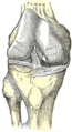 Right knee-joint, from the front, showing interior ligaments.
 -
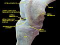
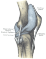 Capsule of right knee-joint (distended). Lateral aspect.
 -
 Capsule of right knee-joint (distended). Posterior aspect.
 -
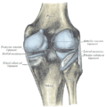
 Anterior view of knee.
 -
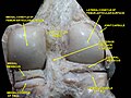 Right knee in extension. Deep dissection. Posterior view.
 -
 Right knee in extension. Deep dissection. Posterior view.
 -
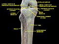 Knee and tibiofibular joint. Deep dissection. Anterior view.
 -
 Knee and tibiofibular joint. Deep dissection. Anterior view.
 -
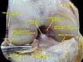
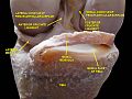
 Knee joint. Deep dissection. Anteromedial view.
 -
 Knee joint. Deep dissection. Anteromedial view.
 -
 Knee joint. Deep dissection. Anteromedial view.
 -
 Knee joint. Deep dissection. Anteromedial view.
 -
 Knee joint. Deep dissection. Anterior view
 -
 Knee joint. Deep dissection. Anterior view.
 -
 Knee joint. Deep dissection. Posterior view.


See also
References
![]() This article incorporates text in the public domain from page 343 of the 20th edition of Gray's Anatomy (1918)
This article incorporates text in the public domain from page 343 of the 20th edition of Gray's Anatomy (1918)
External links
- Anatomy figure: 17:07-09 at Human Anatomy Online, SUNY Downstate Medical Center
- "Anatomy diagram: 02240.009-2". Roche Lexicon - illustrated navigator. Elsevier. Archived from the original on 2012-07-22.
- lljoints at The Anatomy Lesson by Wesley Norman (Georgetown University) (antkneejointopenflexed)