
Posterior cruciate ligament
| Posterior cruciate ligament | |
|---|---|
 Diagram of the knee | |
| Details | |
| From | antero-lateral aspect of medial femoral condyle |
| To | posterolateral aspect of proximal tibia |
| Identifiers | |
| Latin | ligamentum cruciatum posterius genus |
| MeSH | D016119 |
| TA98 | A03.6.08.008 |
| TA2 | 1891 |
| FMA | 44617 |
| Anatomical terminology | |
The posterior cruciate ligament (PCL) is a
. This configuration allows the PCL to resist forces pushing the tibia posteriorly relative to the femur.The PCL and ACL are intracapsular ligaments because they lie deep within the knee joint. They are both isolated from the fluid-filled synovial cavity, with the synovial membrane wrapped around them. The PCL gets its name by attaching to the posterior portion of the tibia.[1]
The PCL, ACL, MCL, and LCL are the four main ligaments of the knee in primates.
Structure
The PCL is located within the

Function
Although each PCL is a unified unit, they are described as separate anterolateral and posteromedial sections based on where each section's attachment site and function.
The function of the PCL is to prevent the femur from sliding off the anterior edge of the tibia and to prevent the tibia from displacing posterior to the femur. The posterior cruciate ligament is located within the knee.
Clinical significance
Common causes of injuries are direct blows to the flexed knee, such as the knee hitting the dashboard in a car accident or falling hard on the knee, both instances displacing the tibia posterior to the femur.[10]
An additional test of posterior cruciate ligament injury is the posterior sag test, where, in contrast to the drawer test, no active force is applied. Rather, the person lies supine with the leg held by another person so that the hip is flexed to 90 degrees and the knee 90 degrees.
There are four different grades of classification in which medical doctors classify a PCL injury:
- Grade I, the PCL has a slight tear.
- Grade II, the PCL ligament is minimally torn and becomes loose.
- Grade III, the PCL is torn completely and the knee can now be categorized as unstable.
- Grade IV, the ligament is damaged along with another ligament housed in the knee (i.e. ACL).
With these grades of PCL injuries, there are different treatments available for such injuries.[citation needed]

Mechanism

In this position, the PCL functions to prevent movement of the tibia in the posterior direction
Treatment
It is possible for the PCL to heal on its own. Even if the PCL does not heal normally, it is unusual for surgery to be required. Treatment is usually physiotherapy to strengthen the muscles around the knee; usually they provide adequate stability even without a functional PCL. Only if there are ongoing symptoms down the track, or if there are other injuries in the knee (e.g. posterolateral corner injury) will ligament reconstruction be required.[18] Ligament reconstruction is used to replace the torn PCL with a new ligament, which is usually a graft taken from the hamstring or Achilles tendon from a host cadaver. An arthroscope allows a complete evaluation of the entire knee joint, including the knee cap (patella), the cartilage surfaces, the meniscus, the ligaments (ACL & PCL), and the joint lining. Then, the new ligament is attached to the bone of the thigh and lower leg with screws to hold it in place.[19] Surgery to repair the posterior cruciate ligament is controversial due to its placement and technical difficulty.[20]
It is possible for the PCL to heal on its own without surgery when it is a Grade I or Grade II injury. PCL injuries that are diagnosed in these categories can have their recovery times reduced by performing certain rehabilitative exercises. Fernandez and Pugh(2012) found that following a PCL grade II diagnosis, a
Other animals
In the
Additional images
-
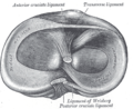 Head of right tibia seen from above, showing menisci and attachments of ligaments.
 -
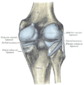 Capsule of right knee-joint (distended). Posterior aspect.
 -
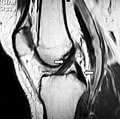 Magnetic resonance imaging evaluation demonstrating normal signal of both anterior and posterior cruciate ligaments (arrows).
 -
 Anterior view of knee.
 -
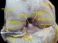
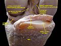 Knee joint. Deep dissection. Anterior view


References
- ^ Saladin, K. S. 2010. Anatomy & Physiology: 5th edition. McGraw-Hill[page needed]
- S2CID 15246937.
- PMID 1126079.
- ^ PMID 23022245.
- PMID 17349472.
- ^ S2CID 3524402.
- ^ PMID 16360655.
- S2CID 42810072.
- PMID 8106532.
- ^ MedlinePlus Encyclopedia: Posterior cruciate ligament (PCL) injury
- ^ Posterior Sag Test From The University of West Alabama, Athletic Training & Sports Medicine Center. Retrieved Feb 2011
- ISBN 0-7216-0013-1.
- S2CID 24386922.
- PMID 23181354.
- ^ S2CID 23746497.
- ISBN 978-0-7817-9128-1.
- ISBN 978-0-7817-9128-1.
- ^ American Academy of Orthopedic Surgeons. "Posterior Cruciate Ligament Injuries". OrthoInfo. American Academy of Orthopedic Surgeons. Retrieved 7 May 2019.
- ^ http://www.orthspec.com/pdfs/PCL-injuries.pdf[full citation needed]
- ^ Jonathan Cluett, M.D. (2003-08-05). "Injuries to the posterior cruciate ligament (PCL)". about.com. Retrieved 2006-11-11.
- PMID 23204951.
- S2CID 22150465.
- ISBN 978-0-7020-2788-8. Retrieved September 8, 2009.
External links
- lljoints at The Anatomy Lesson by Wesley Norman (Georgetown University) (antkneejointopenflexed)
- Dealing with Torn Ligament in the Knee
- http://www.orthspec.com/pdfs/PCL-injuries.pdf