
Lobules of liver
| Lobules of liver | |
|---|---|
 The structure of the liver’s functional units or lobules. Blood enters the lobules through branches of the portal vein and hepatic artery proper, then flows through sinusoids. | |
| Details | |
| System | Digestive system |
| Location | Liver |
| Identifiers | |
| Latin | lobuli hepatis |
| TA98 | A05.8.01.056 |
| TA2 | 3060 |
| FMA | 76488 |
| Anatomical terms of microanatomy | |
In histology (microscopic anatomy), the lobules of liver, or hepatic lobules, are small divisions of the liver defined at the microscopic scale. The hepatic lobule is a building block of the liver tissue, consisting of a portal triad, hepatocytes arranged in linear cords between a capillary network, and a central vein.
Lobules are different from the lobes of liver: they are the smaller divisions of the lobes. The two-dimensional microarchitecture of the liver can be viewed from different perspectives:[1]
| Name | Shape | Model |
| classical lobule[2] | hexagonal; divided into concentric centrilobular, midzonal, periportal parts | anatomical |
| portal lobule[3] | triangular; centered on a portal triad |
bile secretion |
| acinus [4] | elliptical or diamond-shaped; divided into zone I (periportal), zone II (transition zone), and zone III (pericentral) | blood flow and metabolic |
The term "hepatic lobule", without qualification, typically refers to the classical lobule.
Structure
The hepatic lobule can be described in terms of metabolic "zones", describing the hepatic
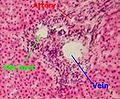
Portal triad
A portal triad (also known as portal canal, portal field[citation needed], portal area[citation needed], or portal tract[citation needed]) is a distinctive arrangement within lobules. It consists of the following five structures:[6]
- proper hepatic artery, an arteriole branch of the hepatic artery that supplies oxygen
- hepatic portal vein, a venule branch of the portal vein, with blood rich in nutrients but low in oxygen
- one or two small bile ductules of cuboidal epithelium, branches of the bile conducting system.
- lymphatic vessels
- branch of the vagus nerve
The misnomer "portal triad" traditionally has included only the first three structures, and was named before lymphatic vessels were discovered in the structure.[7][8] It can refer both to the largest branch of each of these vessels running inside the hepatoduodenal ligament, and to the smaller branches of these vessels inside the liver.
In the smaller portal triads, the four vessels lie in a network of connective tissue and are surrounded on all sides by hepatocytes. The ring of hepatocytes abutting the connective tissue of the triad is called the periportal limiting plate.
-
 Portal triad
 -
 Labeled sketch of a portal canal
 -
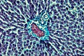 Portal triad of a rat liver, 1 branch of hepatic artery, 2 branch of portal vein, 3 bile duct
 -
 Portal triad of mouse liver. 1= bile duct, 2= branch of hepatic artery, 3= branch of portal vein, 4= lymphatic vessels


.jpg)

Periportal space
The periportal space (
Fluid (residual blood plasma) that is not taken up by hepatocytes drains into the periportal space, and is taken up by the lymphatic vessels that accompany the other
Function

Zones differ by function:
- zone I hepatocytes are specialized for oxidative liver functions such as cholesterol synthesis
- zone III cells are more important for acetaminophen toxicity.[12]
Other zonal injury patterns include zone I deposition of
Clinical significance
Bridging fibrosis, a type of fibrosis seen in several types of liver injury, describes fibrosis from the central vein to the portal triad.[13]
See also
References
- ^ Cell and Tissue Structure at U. Va.
- ^ Histology image: 88_03 at the University of Oklahoma Health Sciences Center
- ^ Histology image: 88_09a at the University of Oklahoma Health Sciences Center
- ^ Histology image: 88_09b at the University of Oklahoma Health Sciences Center
- ISBN 0-323-03675-9.
- ISBN 978-0-07-180720-3.
- ISBN 9781478436232.
- ^ "Physiology of the Hepatic Vascular System". www.vivo.colostate.edu. Retrieved 2018-12-04.
- ISBN 978-0-443-10012-3. Retrieved 23 May 2011.
- ISBN 0-7817-7221-4.
- ^ ISBN 978-0-7817-6040-9.
- ^ M.J. Burns; S.L. Friedman; A.M. Larson (2009). "Acetaminophen (paracetamol) poisoning in adults: Pathophysiology, presentation, and diagnosis". In D.S. Basow (ed.). UpToDate. Waltham, MA: UpToDate.
- ^ "The liver ~ Medical student education – Tissupath". tissupath.com.au. Retrieved 20 June 2018.
External links
- Histology at siumed.edu
- Histology at okstate.edu
- UIUC Histology Subject 923