Protocadherin
| Protocadherin, cytoplasmic | |||||||||
|---|---|---|---|---|---|---|---|---|---|
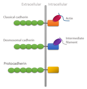
 Domain organization of different types of cadherins showing unique features of protocadherins: Extracellular domain is longer and intracellular domain lacks attachment with cytoskeleton. | |||||||||
| Identifiers | |||||||||
| Symbol | PDCH | ||||||||
| Pfam | PF08374 | ||||||||
| InterPro | IPR013585 | ||||||||
| Membranome | 114 | ||||||||
| |||||||||
Protocadherins (Pcdhs) are the largest mammalian subgroup of the
Classification
In mammals, two types of Pcdh genes have been defined: the non-clustered Pcdhs which are scattered throughout the genome; and the clustered Pcdhs organized in three gene clusters designated α, β, γ which in mouse genome comprises 14, 22 and 22, respectively, large variable exons arrayed in tandem. Each exon is transcribed from its owner promoter and encodes: the entire extracellular domain, a transmembrane domain, and a short and variable intracellular domain of the corresponding Pcdh protein which differs from the Cadherin intracellular domain due to lack of attachment to the cytoskeleton through catenins.[5]
Moreover, these clustered Pcdh genes are predominantly expressed in the developing nervous system[2] and since different subsets of Pcdhs genes are differentially expressed in individual neurons, a vast cell surface diversity may arise from this combinatorial expression.[5] This has led to speculation and further to the proposal that Pcdhs may provide a synaptic-address code for neuronal connectivity or a single-cell barcode for self-recognition/self-avoidance similar to that ascribed to DSCAM proteins of invertebrates. Although vertebrate DSCAMs lack the diversity of their invertebrate counterparts, the selective transcription of individual Pcdh isoforms can be achieved by promoter choice followed by alternative pre-mRNA cis-splicing thus increasing the number of possible combinations.
Function
Homophilic interactions and intracellular signaling
Clustered Pcdhs proteins are detected throughout the neuronal soma, dendrites and axons and are observed in synapses and growth cones.[6][7][8][9][10] Like classical cadherins, members of Pcdhs family were also shown to mediate cell-cell adhesion in cell-based assays[11][12][13] and most of them showed to engage in homophilic trans-interactions.[14] Schreiner and Weiner [14] showed that Pcdhα and γ proteins can form multimeric complexes. If all three classes of Pcdhs could engage in multimerization of stochastically expressed Pcdhs isoforms, then neurons could produce a large number of distinct homophilic interaction units, amplifying significantly the cell-surface diversity more than the one afforded by stochastic gene expression alone.
As for cytoplasmic domain, all the three classes of clustered Pcdhs proteins are dissimilar, although they are strictly conserved in vertebrate evolution, suggesting a conserved cellular function.
The cytoplasmic domain of Pcdh-alpha can be divided into two specific types. Both of them enhance homophilic interactions. They associate with neurofillament M and fascin respectively.[22]
Human genes
- PCDH1
- PCDH7
- PCDH8
- PCDH9
- PCDH10
- PCDH11X/11Y
- PCDH12
- PCDH15
- PCDH17
- PCDH18
- PCDH19
- PCDH20
- PCDHA1
- PCDHA2
- PCDHA3
- PCDHA4
- PCDHA5
- PCDHA6
- PCDHA7
- PCDHA8
- PCDHA9
- PCDHA10
- PCDHA11
- PCDHA12
- PCDHA13
- PCDHAC1
- PCDHAC2
- PCDHB1
- PCDHB2
- PCDHB3
- PCDHB4
- PCDHB5
- PCDHB6
- PCDHB7
- PCDHB8
- PCDHB9
- PCDHB10
- PCDHB11
- PCDHB12
- PCDHB13
- PCDHB14
- PCDHB15
- PCDHB16
- PCDHB17
- PCDHB18
- PCDHGA1
- PCDHGA2
- PCDHGA3
- PCDHGA4
- PCDHGA5
- PCDHGA6
- PCDHGA7
- PCDHGA8
- PCDHGA9
- PCDHGA10
- PCDHGA11
- PCDHGA12
- PCDHGB1
- PCDHGB2
- PCDHGB3
- PCDHGB4
- PCDHGB5
- PCDHGB6
- PCDHGB7
- PCDHGC3
- PCDHGC4
- PCDHGC5
- FAT
- FAT2
- FAT4
See also
- Cadherin
- PCDH11X
- Neuronal self-avoidance
- Epileptic Encephalopathy, Early Infantile, 9, caused by mutation in the gene encoding protocadherin-19
References
- PMID 18848899.
- ^ PMID 8508762.
- ^ PMID 22884324.
- PMID 26268193.
- ^ PMID 23900538.
- PMID 9655502.
- PMID 12467588.
- S2CID 41159576.
- PMID 12832533.
- S2CID 24229102.
- PMID 8719883.
- S2CID 40074917.
- PMID 16751190.
- ^ PMID 20679223.
- PMID 20616001.
- PMID 19136062.
- PMID 17403907.
- PMID 20876099.
- PMID 11007549.
- S2CID 26590444.
- S2CID 11421820.
- PMID 16408303.
Further reading
- Han MH, Lin C, Meng S, Wang X (January 2010). "Proteomics analysis reveals overlapping functions of clustered protocadherins". Molecular & Cellular Proteomics. 9 (1): 71–83. PMID 19843561.
- Sotomayor M, Gaudet R, Corey DP (September 2014). "Sorting out a promiscuous superfamily: towards cadherin connectomics". Trends in Cell Biology. 24 (9): 524–36. PMID 24794279.
- Pancho A, Aerts T, Mitsogiannis MD, Seuntjens E (June 2020). "PProtocadherins at the Crossroad of Signaling Pathways". Frontiers in Molecular Neuroscience. 13: 117. PMID 32694982.
