
Supraspinous fossa
| Supraspinous fossa | |
|---|---|
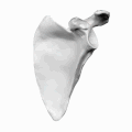
 Left scapula. Dorsal surface. Supraspinatous fossa shown in red. | |
 Left scapula. Dorsal surface. Supraspinatous fossa shown in red. | |
| Details | |
| Identifiers | |
| Latin | fossa supraspinata |
| TA98 | A02.4.01.007 |
| TA2 | 1150 |
| FMA | 23269 |
| Anatomical terms of bone | |
The supraspinous fossa (supraspinatus fossa, supraspinatous fossa) of the posterior aspect of the
Structure
The fossa can be exposed by the removal of skin and the
The supraspinous fossa is bounded by the
Supraspinatus muscle originates from the supraspinous fossa. Distal attachment of the levator scapulae muscle is also on the medial aspect of the fossa.
Function
The
Clinical significance
Rotator cuff tear
Hollowing in the supraspinous and the infraspinous area is frequently seen as chronic rotator cuff tear resulting in wasting.[2] The wasting may be caused by the supraglenoid cyst compressing the suprascapular nerve and causes a loss of innervation to supraspinatus and infraspinatus muscles. Such wasting or hollowing can be differentially diagnosed as nerve compression or tendon rupture.
Additional images
-
 The human scapula
 -
 Supraspinous fossa shown in red.
 -
 Supraspinous fossa shown in red.


See also
References
- ISBN 9781451119459.
- ISBN 9780736086431.
![]() This article incorporates text in the public domain from page 203 of the 20th edition of Gray's Anatomy (1918)
This article incorporates text in the public domain from page 203 of the 20th edition of Gray's Anatomy (1918)
External links
- Anatomy figure: 03:01-02 at Human Anatomy Online, SUNY Downstate Medical Center