
Ulnar artery
This article about biology may be excessively human-centric. (October 2017) |
| Ulnar artery | |
|---|---|
Ulnar vein | |
| Identifiers | |
| Latin | arteria ulnaris |
| MeSH | D017535 |
| TA98 | A12.2.09.041 |
| TA2 | 4655 |
| FMA | 22796 |
| Anatomical terminology] | |
The ulnar artery is the main
Along its course, it is accompanied by a similarly named
The ulnar artery, the larger of the two terminal branches of the brachial, begins a little below the bend of the
Branches
Forearm: Anterior ulnar recurrent artery, Posterior ulnar recurrent artery,
Hand: Deep palmar branch of ulnar artery which passes through the hypothenar muscles to anastomose with the deep palmar arch which is formed predominantly by the radial artery and the terminal branch of the ulnar artery is then to form the superficial palmar arch.
Relations
In its upper half, it is deeply seated, being covered by the
The
In the lower half of the forearm it lies upon the
It is accompanied by two
Wrist
At the wrist the ulnar artery is covered by the integument and the
Peculiarities
The ulnar artery varies in its origin in the proportion of about one in thirteen cases; it may arise about 5 to 7 cm. below the elbow, but more frequently higher, the brachial being more often the source of origin than the axillary.
Variations in the position of this vessel are more common than in the radial. When its origin is normal, the course of the vessel is rarely changed.
When it arises high up, it is almost invariably superficial to the Flexor muscles in the forearm, lying commonly beneath the fascia, more rarely between the fascia and integument.
In a few cases, its position is subcutaneous in the upper part of the forearm, and subaponeurotic in the lower part.
See also
- Allen test
Additional images
-
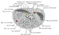
 Cross-section through the middle of the forearm.
 -
 Transverse section across distal ends of radius and ulna.
 -
 Transverse section across the wrist and digits.
 -
 The palmar aponeurosis.
 -
 Diagram of the anastomosis around the elbow-joint.
 -
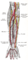
 Arteries of the right forearm - anterior view.
 -
 Ulnar and radial arteries. Deep view.


References
![]() This article incorporates text in the public domain from page 595 of the 20th edition of Gray's Anatomy (1918)
This article incorporates text in the public domain from page 595 of the 20th edition of Gray's Anatomy (1918)
External links
- Ulnar artery at the Duke University Health System's Orthopedics program
- lesson4artofforearm at The Anatomy Lesson by Wesley Norman (Georgetown University)