
Circle of Willis
| Circle of Willis | |
|---|---|
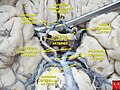
 Diagram of the arterial circulation at the base of the brain (inferior view), the circle of Willis is drawn in the upper half. Blood flows up to the brain through the vertebral arteries and through the internal carotid arteries. | |
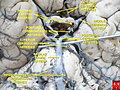
 The arteries of the base of the brain. Basilar artery labeled below center. The temporal pole of the cerebrum and the cerebellar hemisphere have been removed on the right side. Inferior aspect (viewed from below). | |
| Details | |
| Identifiers | |
| Latin | Circulus arteriosus cerebri Circulus Willisii |
| MeSH | D002941 |
| TA98 | A12.2.07.080 |
| TA2 | 4516 |
| FMA | 50454 |
| Anatomical terminology | |
The circle of Willis (also called Willis' circle, loop of Willis, cerebral arterial circle, and Willis polygon) is a circulatory anastomosis that supplies blood to the brain and surrounding structures in reptiles, birds and mammals, including humans.[1] It is named after Thomas Willis (1621–1675), an English physician.[2]
Structure
The circle of Willis is a part of the cerebral circulation and is composed of the following arteries:[3]
- Anterior cerebral artery (left and right) at their A1 segments
- Anterior communicating artery
- Internal carotid artery (left and right) at its distal tip (carotid terminus)
- Posterior cerebral artery (left and right) at their P1 segments
- Posterior communicating artery (left and right)
The
Origin of arteries
The left and right internal carotid arteries arise from the left and right common carotid arteries.
The posterior communicating artery is given off as a branch of the internal carotid artery just before it divides into its terminal branches - the anterior and middle cerebral arteries. The anterior cerebral artery forms the anterolateral portion of the circle of Willis, while the middle cerebral artery does not contribute to the circle.
The right and left posterior cerebral arteries arise from the basilar artery, which is formed by the left and right vertebral arteries. The vertebral arteries arise from the subclavian arteries.
The anterior communicating artery connects the two anterior cerebral arteries and could be said to arise from either the left or right side.
All arteries involved give off cortical and central branches. The central branches supply the interior of the circle of Willis, more specifically, the Interpeduncular fossa. The cortical branches are named for the area they supply and do not directly affect the circle of Willis.
Variation
Considerable
Function
The arrangement of the brain's arteries into the circle of Willis is believed to create redundancy (analogous to engineered redundancy) for collateral circulation in the cerebral circulation. If one part of the circle becomes blocked or narrowed (stenosed) or one of the arteries supplying the circle is blocked or narrowed, blood flow from the other blood vessels can often preserve the cerebral perfusion well enough to avoid the symptoms of ischemia.[6]
However, considering that the circle of Willis is present in many non-human species (reptiles, birds and mammals), and that arterial narrowing is mostly associated with old age and the human lifestyle, more generally applicable explanations of its functions have been suggested, such as dampening of pulse pressure waves within the brain[7] and involvement in forebrain sensing of water loss.[1]
Clinical significance
Aneurysms

Subclavian steal syndrome
The adaptive flow that the circle of Willis introduces can also lead to reduced cerebral perfusion.[8][9] In subclavian steal syndrome, blood is "stolen" from the vertebral artery on the affected side to preserve blood flow to the upper limb. Subclavian steal syndrome results from a proximal stenosis (narrowing) of the subclavian artery, one of arteries originating off of the aortic arch. Subclavian steal syndrome has potential to affect flow in the circle of Willis.
Additional images
-
 Fetal ultrasound image at the level of circle of Willis, showing PCA, MCA and ACA
 -
 Cerebralposterior cerebralcirculation, the posterior aspect of the circle of Willis, and one of its feeding vessels
 -
 An anterior view of major cerebral and cerebellar arteries.
 -

-
 Circle of Willis
 -
 Circle of Willis


See also
References
- ^ PMID 33191609.
- S2CID 146301989.
- ]
- ^ Bergman, Ronald A.; Afifi, Adel K.; Miyauchi, Ryosuke (2005). "Circle of Willis". Illustrated Encyclopedia of Human Anatomic Variation: Opus II: Cardiovascular System: Arteries: Head, Neck, and Thorax.
- S2CID 218989565.
- S2CID 22591168.
- PMID 24473483.
- PMID 3041649.
- PMID 5377222.
External links
- Bergman, Ronald A.; Afifi, Adel K.; Miyauchi, Ryosuke. "Fourteen Variations of Circle of Willis and Related Vessels". Illustrated Encyclopedia of Human Anatomic Variation: Opus II: Cardiovascular System.