Dynamin
| Dynamin family | |||||||||
|---|---|---|---|---|---|---|---|---|---|
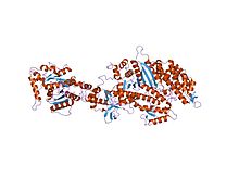
 Structure of the nucleotide-free myosin II motor domain from Dictyostelium discoideum fused to the GTPase domain of dynamin I from Rattus norvegicus | |||||||||
| Identifiers | |||||||||
| Symbol | Dynamin_N | ||||||||
| Pfam | PF00350 | ||||||||
| Pfam clan | CL0023 | ||||||||
| InterPro | IPR001401 | ||||||||
| PROSITE | PDOC00362 | ||||||||
| |||||||||
| Dynamin central region | |||||||||
|---|---|---|---|---|---|---|---|---|---|
 Structure of the nucleotide-free myosin II motor domain from Dictyostelium discoideum fused to the GTPase domain of dynamin I from Rattus norvegicus | |||||||||
| Identifiers | |||||||||
| Symbol | Dynamin_M | ||||||||
| Pfam | PF01031 | ||||||||
| InterPro | IPR000375 | ||||||||
| |||||||||
Dynamin is a
Structure

Dynamin itself is a 96
Function
During clathrin-mediated endocytosis, the cell membrane invaginates to form a budding vesicle. Dynamin binds to and assembles around the neck of the endocytic vesicle, forming a helical polymer arranged such that the GTPase domains dimerize in an asymmetric manner across helical rungs.[7][8] The polymer constricts the underlying membrane upon GTP binding and hydrolysis via conformational changes emanating from the flexible neck region that alters the overall helical symmetry.[8] Constriction around the vesicle neck leads to the formation of a hemi-fission membrane state that ultimately results in membrane scission.[2][6][9] Constriction may be in part the result of the twisting activity of dynamin, which makes dynamin the only molecular motor known to have a twisting activity.[10]
Types
In mammals, three different dynamin genes have been identified with key sequence differences in their Pleckstrin homology domains leading to differences in the recognition of lipid membranes:
- Dynamin I is expressed in neurons and neuroendocrine cells
- Dynamin II is expressed in most cell types
Pharmacology
Small molecule inhibitors of dynamin activity have been developed, including Dynasore[11][12] and photoswitchable derivatives (Dynazo)[13] for spatiotemporal control of endocytosis with light (photopharmacology).
Disease implications
Mutations in
References
- ^ S2CID 24401725.
- ^ a b Hinshaw, J. "Research statement, Jenny E. Hinshaw, Ph.D." Archived 2021-07-15 at the Wayback Machine National Institute of Diabetes & Digestive & Kidney Diseases, Laboratory of Cell Biochemistry and Biology. Accessed 19 March 2013.
- ^ PMID 9012790.
- PMID 16218949.
- S2CID 4365628.
- ^ S2CID 6305282.
- PMID 25088425.
- ^ PMID 30069048.
- PMID 26123023.
- S2CID 4413887.
- PMID 16740485.
- S2CID 51895475.
- PMID 33016959.
- S2CID 19191581.
- PMID 27066543.
External links
- Dynamins at the U.S. National Library of Medicine Medical Subject Headings (MeSH)
