
Hemangioblastoma
| Hemangioblastoma | |
|---|---|
cerebellar hemangioblastoma. HPS stain. | |
| Specialty | Oncology |
Hemangioblastomas, or haemangioblastomas, are
Presentation
Complications
Hemangioblastomas can cause an abnormally high number of red blood cells in the bloodstream due to ectopic production of the hormone erythropoietin as a paraneoplastic syndrome.[3]
Pathogenesis
Hemangioblastomas are composed of
Diagnosis

The primary diagnosis is made with a
Treatment
The treatment for hemangioblastoma is
Prognosis
The outcome for hemangioblastoma is very good if surgical extraction of the tumor can be achieved; excision is possible in most cases and permanent neurologic deficit is uncommon and can be avoided altogether if the tumor is diagnosed and treated early. Persons with VHL syndrome have a bleaker prognosis than those who have sporadic tumors since those with VHL syndrome usually have more than one lesion.[2]
Epidemiology
Hemangioblastoma are among the rarest central nervous system tumors, accounting for less than 2%. Hemangioblastomas usually occur in adults, yet tumors may appear in VHL syndrome at much younger ages. Men and women are approximately at the same risk. Although they can occur in any section of the
Additional images
-
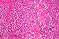 Micrograph of cerebellar hemangioblastoma. HPS stain.
 -
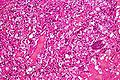 Micrograph of cerebellar hemangioblastoma. HPS stain.


References
- ^ ISBN 0-443-04345-0.
- ^ ISBN 92-832-2430-2.
- PMID 8775737.
- .
- ^ "Hemangioblastoma". The Lecturio Medical Concept Library. Retrieved 24 July 2021.
- S2CID 19652430.
- S2CID 20823909.