Hypophysitis
| Hypophysitis | |
|---|---|
 | |
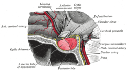
| Pituitary gland is located at the base of the human brain. | |
| Specialty | Endocrinology |
Hypophysitis refers to an inflammation of the pituitary gland. Hypophysitis is rare and not fully understood.
Signs and symptoms
There are four categories of symptoms and signs. Most commonly, the initial symptoms are headaches and visual disturbances. Some symptoms are derived from the lesser functioning of the adenohypophyseal hormones. Of the adenohypophyseal hormones, the most frequently affected are
Cause
Hypophysitis may have an underlying autoimmune aetiology, as in the case of autoimmune hypophysitis,[2] and lymphocytic hypophysitis.[3][4]
Diagnosis
Mainly, the diagnosis of hypophysitis is through exclusion – patients often undergo surgery because they are suspected of having a pituitary adenoma. But, the most accurate diagnosis is using Magnetic Resonance Imaging (MRI) to find any mass or lesions on the sella turcica.
Treatment
It was shown through various testing that administration of
Prognosis
The prognosis for hypophysitis was variable for each individual. The depending factors for hypophysitis included the advancement of the mass on the sella turcica, percentage of fibrosis, and the body's response to corticosteroids. Through the use of corticosteroids, the vision defects tend to recover when the gland size began to decrease. The prognoses of the limited number of reported cases were usually good.[5]
History
The first reported case was in 1962, with a 22-year-old who died of adrenal insufficiency 14 months after giving birth to her second child. Her symptoms began 3 months postpartum, with
See also
- Autoimmune hypophysitis[2]
- Lymphocytic hypophysitis[3][4]
- Pituitary disease
- Hypopituitarism
References
- ^ "Autoimmune Hypophysitis Symptoms". Johns Hopkins University School of Medicine & Johns Hopkins Health System. 2002. Archived from the original on 2004-05-28.
- ^ PMID 17554056.
- ^ PMID 7629223.
- ^ a b "Lymphoid Hypophisitis". pituitaryadenomas.com. Archived from the original on 2020-10-21. Retrieved 2011-11-10.
- ^ a b c "Unknown". Archived from the original on 2016-03-04. Retrieved 2011-11-11.
- PMID 11238484.