
Sella turcica
| Sella turcica | |
|---|---|
 Human skull seen from side (parietal bones and temporal bones have been removed). Sella turcica shown in red. | |
 Sella turcica and pituitary gland. | |
| Details | |
| Identifiers | |
| Latin | sella turcica |
| MeSH | D012658 |
| TA98 | A02.1.05.006 |
| TA2 | 589 |
| FMA | 54709 |
| Anatomical terms of bone | |
The sella turcica (
Structure
The sella turcica is located in the sphenoid bone behind the chiasmatic groove and the tuberculum sellae. It belongs to the middle cranial fossa.[1]
The sella turcica's most inferior portion is known as the hypophyseal fossa (the "seat of the saddle"), and contains the pituitary gland (hypophysis). In front of the hypophyseal fossa is the tuberculum sellae.
Completing the formation of the saddle posteriorly is the dorsum sellae, which is continuous with the clivus, inferoposteriorly. The dorsum sellae is terminated laterally by the posterior clinoid processes.
Development
It is widely believed that the development of the diaphragma sellae is a factor which determines the morphology of the sella turcica and its contents.[2]
Function
The sella turcica forms a bony seat for the pituitary gland.
Clinical significance
Empty sella syndrome is the condition of a shrunken or flattened pituitary gland.
Since the sella turcica forms a bony
Some pituitary adenomas can extend inferiorly, growing downward and invading the sphenoid bone and cavernous sinus.[3] Large adenomas can cause remodeling of the underlying sphenoid bone altering the shape of the sella turcica.[citation needed]
Sella turcica is also usually used as a reference point with
Etymology
Sella turcica is from the Latin words sella, meaning seat, and turcica, meaning Turkish.
See also
- Sphenoidal sinuses
- Empty sella syndrome
Additional images
-
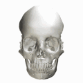 Human skull seen from side (parietal bones and temporal bones have been removed). Sella turcica shown in red.
 -
 Hypophysial fossa shown in red.
 -
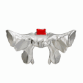 Sphenoid bone seen from above. Sella turcica shown in red.
 -
 Base of skull - Sella turcica, tuberculum sellae and hypophyseal fossa


References
- ISBN 9781437735802.
- S2CID 32329369.
- PMID 8232800.
- Proffit, William R.Contemporary Orthodontics, 4th Edition. C.V. Mosby, 122006. 6.5.2.1). vbk:978-0-323-04046-4#outline(6.5.2.1)
- Marieb, Elaine Nicpon (2004). Human Anatomy & Physiology (6th ed.). Pearson Education. p. 209. ISBN 0-8053-5462-X.