
Teres minor muscle
| Teres minor muscle | |
|---|---|
Laterally rotates the arm, stabilizes humerus | |
| Identifiers | |
| Latin | musculus teres minor |
| TA98 | A04.6.02.010 |
| TA2 | 2459 |
| FMA | 32550 |
| Anatomical terms of muscle] | |
The teres minor (Latin teres meaning 'rounded') is a narrow, elongated muscle of the rotator cuff. The muscle originates from the lateral border and adjacent posterior surface of the corresponding right or left scapula and inserts at both the greater tubercle of the humerus and the posterior surface of the joint capsule.[1]
The primary function of the teres minor is to modulate the action of the deltoid, preventing the humeral head from sliding upward as the arm is abducted. It also functions to rotate the humerus laterally. The teres minor is innervated by the axillary nerve.[2]
Structure
It arises from the dorsal surface of the axillary border of the
Its fibers run obliquely upwards and laterally; the upper ones end in a tendon which is inserted into the lowest of the three impressions on the greater tubercle of the humerus; the lowest fibers are inserted directly into the humerus immediately below this impression.
Relations
The teres minor originates at the lateral border and adjacent posterior surface of the
Innervation
The muscle is innervated by the posterior branch of axillary nerve where it forms a
Variation
Sometimes a group of muscle fibres from teres minor may be fused with
Function
The
Clinical significance
Injury
There are two types of rotator cuff injuries: acute tears and chronic tears. Acute tears occur as a result of a sudden movement. This might include throwing a powerful pitch, holding a fast moving rope during water sports, falling over onto an outstretched hand at speed, or making a sudden thrust with the paddle in kayaking. A chronic tear develops over a period of time. They usually occur at or near the tendon, as a result of the tendon rubbing against the underlying bone.[4] The teres minor is typically normal following a rotator cuff tear.[5]
Imaging


Additional images
-
 Position of the teres minor muscles (shown in red). Animation.
 -
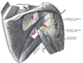 Suprascapular and axillary nerves of right side, seen from behind. (Teres minor is visible at center.)
 -
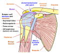 Diagram of the human shoulder joint, front view
 -
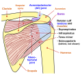 Diagram of the human shoulder joint, back view
 -
 Left scapula. Dorsal surface.
 -
 Left humerus. Posterior view.
 -
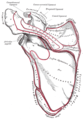
 The scapular and circumflex arteries.
 -
 The suprascapular, axillary, and radial nerves.
 -
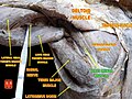 Teres minor muscle


See also
- Accessory muscles of the scapula
References
![]() This article incorporates text in the public domain from page 441 of the 20th edition of Gray's Anatomy (1918)
This article incorporates text in the public domain from page 441 of the 20th edition of Gray's Anatomy (1918)
- ISBN 9780073403717.
- ISBN 9780073403717.
- PMID 13475146.
- ISBN 0-7360-4117-6.
- S2CID 8639793. Retrieved 28 November 2016.
- S2CID 6341877.
External links
- Anatomy figure: 03:03-05 at Human Anatomy Online, SUNY Downstate Medical Center
- ExRx