
Supinator muscle
| Supinator muscle | |
|---|---|
pronator quadratus | |
| Identifiers | |
| Latin | musculus supinator |
| TA98 | A04.6.02.048 |
| TA2 | 2512 |
| FMA | 38512 |
| Anatomical terms of muscle] | |
In human anatomy, the supinator is a broad muscle in the
Structure
The supinator consists of two planes of fibers, between which passes the
The superficial fibers (pars superficialis) surround the upper part of the radius, and are inserted into the lateral edge of the
The proximal aspect of the superficial head is known as the arcade of Frohse or the supinator arch.
Innervation
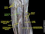
It is innervated by the deep branch of the radial nerve. The deep branch then becomes the posterior interosseous nerve upon exiting the supinator muscle. Its nerve roots are primarily from C6, with some C5 involvement. There is also possible additional C7 innervation.
The radial nerve divides into deep and sensory superficial branches just proximal to the supinator muscle—an arrangement that can lead to entrapment and compression of the deep part, potentially resulting in selective paralysis of the muscles served by this nerve (the extensor muscles and the abductor pollicis longus.)[3] Many possible causes are known for this nerve syndrome, known as supinator entrapment syndrome, including compression by various soft-tissued masses surrounding the nerve, and stress caused by repetitive supination and pronation.[4]
Variation
The deep radial nerve passes through the belly of supinator in 70% of cases and via the arcade of Frohse in remaining cases.[5]
Function
Encircling the radius, supinator brings the hand into the supinated position. In contrast to the
Supinator always acts together with biceps, except when the elbow joint is extended.[6] It is the most active muscle in forearm supination during unresisted supination, while biceps becomes increasingly active with heavy loading. Supination strength decreases by 64% if supinator is disabled by, for example, injury.[7]
History
Etymology
The term "supinator" can also refer more generally to a muscle that causes supination of a part of the body. In older texts, the term "supinator longus" was used to refer to the brachioradialis, and "supinator brevis" was used to describe the muscle now known as the supinator.
Additional images
-
 Bones of left forearm. Anterior aspect.
 -
 Bones of left forearm. Posterior aspect.
 -
 Front of the left forearm. Deep muscles.
 -
 Posterior surface of the forearm. Deep muscles.
 -
 Ulnar and radial arteries. Deep view.
 -
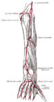 Arteries of the back of the forearm and hand.
 -
 Supinator muscle
 -
 Muscles of upper limb. Cross section.
 -
 Elbow joint. Deep dissection. Anterior view.


.jpg)
.jpg)
Notes
![]() This article incorporates text in the public domain from page 454 of the 20th edition of Gray's Anatomy (1918)
This article incorporates text in the public domain from page 454 of the 20th edition of Gray's Anatomy (1918)
- ^ a b c Platzer 2004, p. 168
- ^ ISBN 978-1-61640-469-7.
- ^ Ross & Lamperti 2006, p. 345
- ^ Chien et al. 2003, Discussion
- ^ Boles, Kannam & Cardwell 2000, p. 153
- ^ "Supinator". Loyola University Medical Education Network. Retrieved 22 March 2011.
- ^ Duqion, Chavan & Bisson 2010, p. 414
References
- Boles, CA; Kannam, S; Cardwell, AB (2000). "The Forearm: Anatomy of Muscle Compartments and Nerves". Am. J. Roentgenol. 174 (1): 151–159. PMID 10628472.
- Chien, A; Jamadar, DA; Jacobson, JA; Hayes, CW; Louis DS (2003). "Sonography and MR Imaging of Posterior Interosseous Nerve Syndrome with Surgical Correlation". Am. J. Roentgenol. 181 (1): 219–221. PMID 12818863.
- Duqion, TR; Chavan, RC; Bisson, LJ (2010). "Innervation of the Supinator Muscle and Its Relationship to Two-Incision Distal Biceps Tendon Repair: An Anatomic Study" (PDF). Clinical Anatomy. 23 (4): 413–419. S2CID 21578840.
- Platzer, W (2004). Color Atlas of Human Anatomy, Vol. 1: Locomotor System (5th ed.). Thieme. ISBN 1-58890-159-9.
- Ross, Lawrence M.; Lamperti, Edward D., eds. (2006). Thieme Atlas of Anatomy: General Anatomy and Musculoskeletal System. Thieme. ISBN 1-58890-419-9.