
Extensor pollicis longus muscle
| Extensor pollicis longus muscle | |
|---|---|
| Identifiers | |
| Latin | musculus extensor pollicis longus |
| TA98 | A04.6.02.051 |
| TA2 | 2516 |
| FMA | 38521 |
| Anatomical terms of muscle] | |
In
Structure
The extensor pollicis longus arises from the dorsal surface of the
Passing through the third tendon compartment,[1] lying in a narrow, oblique groove on the back of the lower end of the radius,[3] it crosses the wrist close to the dorsal midline before turning towards the thumb using Lister's tubercle on the distal end of the radius as a pulley.[2]
It obliquely crosses the tendons of the
At the proximal phalanx, the tendon is joined by expansions from abductor pollicis brevis and adductor pollicis.[2]
The tendon is finally inserted on the base of the
6.7 to 9.7 centimetres (2.6 to 3.8 in) in length, the tendon passes through a long and superficial
Relations
Together with the tendons of the extensor pollicis brevis and the abductor pollicis longus, its tendon crosses the radial artery.[3]
Blood supply
The tendon of extensor pollicis longus is supplied by branches from various arteries. Before the tendon enters its synovial sheath, arteries from the
Innervation
The extensor pollicis longus muscle receives innervation from the posterior interosseous nerve (C7 and C8) which is the continuation of the deep branch of the radial nerve.
Function
Extensor pollicis longus extends the terminal phalanx of the thumb. While abductor pollicis brevis and adductor pollicis, both attached to the extensor pollicis longus tendon, can extend the thumb's interphalangeal joint to the neutral position, only extensor pollicis longus can achieve full hyperextension at the interphalangeal joint. This complete extension at the interphalangeal joint is not possible, or considerably more difficult, with the carpal, carpometacarpal, and metacarpophalangeal joints simultaneously extended. Likewise, flexion at the interphalangeal joint by flexor pollicis longus is considerably reduced in wrist flexion.[2]
It also applies an extensor force at the metacarpophalangeal joint together with the extensor pollicis brevis and extends and adducts at the carpometacarpal joint of the thumb.[2]
Clinical significance
Injury
Tenosynovitis, inflammatory irritation of the synovial sheath, is relatively common in the third compartment after repetitive activities such as drum playing.[5]
Additional images
-
 Anatomical snuff box
 -
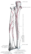 Bones of left forearm. Posterior aspect.
 -
 Bones of the left hand. Dorsal surface.
 -
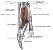 Tendons of forefinger and vincula tendina.
 -
 Cross-section through the middle of the forearm.
 -
 Posterior surface of the forearm. Superficial muscles.
 -
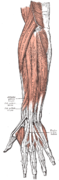
 Posterior surface of the forearm. Deep muscles.
 -
 Transverse section across the wrist and digits.
 -
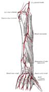 Arteries of the back of the forearm and hand.
 -
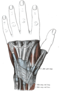 The mucous sheaths of the tendons on the back of the wrist. (Extensor pollicis longus visible at center right.)
 -
 Muscle of the hand . Posterior view.


Notes
![]() This article incorporates text in the public domain from page 455 of the 20th edition of Gray's Anatomy (1918)
This article incorporates text in the public domain from page 455 of the 20th edition of Gray's Anatomy (1918)
- ^ a b c Platzer 2004, p. 168
- ^ a b c d e Austin 2005, p. 339
- ^ a b c Gray's Anatomy 1918, see infobox
- ^ a b Zbrodowski, Gajisin & Grodecki 1982, Results, pp. 235–9
- ^ Schmitt, Lanz & Buchberger 2008, p. 336
References
- Austin, Noelle M (2005). "Chapter 9: The Wrist and Hand Complex". In Levangie, Pamela K; Norkin, Cynthia C (eds.). Joint Structure and Function: A Comprehensive Analysis (4th ed.). Philadelphia: F. A. Davis Company. ISBN 0-8036-1191-9.
- Platzer, Werner (2004). Color Atlas of Human Anatomy, Vol. 1: Locomotor System (5th ed.). ISBN 3-13-533305-1.
- Schmitt, Rainer; Lanz, Ulrich; Buchberger, Wolfgang (2008). Diagnostic Imaging of the Hand. Thieme. ISBN 9781588904539.
- Zbrodowski, A; Gajisin, S; Grodecki, J (September 1982). "Vascularization of the tendons of the extensor pollicis longus, extensor carpi radialis longus and extensor carpi radialis brevis muscles". J Anat. 135 (Pt 2): 235–44. PMID 7174499.