
Teres major muscle
| Teres major muscle | |
|---|---|
Internal rotation (medial rotation) of the humerus, extend the humerus from flexed position | |
| Identifiers | |
| Latin | musculus teres major |
| TA98 | A04.6.02.011 |
| TA2 | 2462 |
| FMA | 32549 |
| Anatomical terms of muscle] | |
The teres major muscle is a muscle of the upper limb. It attaches to the scapula and the humerus and is one of the seven scapulohumeral muscles. It is a thick but somewhat flattened muscle.
The teres major muscle (from Latin teres, meaning "rounded") is positioned above the latissimus dorsi muscle and assists in the extension and medial rotation of the humerus. This muscle is commonly confused as a rotator cuff muscle, but it is not because it does not attach to the capsule of the shoulder joint, unlike the teres minor muscle for example.
Structure
The teres major muscle originates on the dorsal surface of the inferior angle and the lower part of the lateral border of the scapula.
The fibers of teres major insert into the medial lip of the intertubercular sulcus of the humerus.
Relations
The
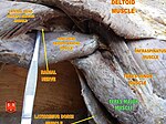
Together with teres minor muscle, teres major muscle forms the axillary space, through which several important arteries and veins pass.[1][2]
Innervation
Teres major is supplied primarily by the lower subscapular nerve[3] and additionally by the thoracodorsal nerve (middle subscapular nerve). These are distal to the upper subscapular nerve. These three nerves branch off the posterior cord of the brachial plexus. The nerves that innervate teres major consist of fibers from spinal nerves C5-C8.[3]
Function
The teres major is a medial
Injury
Isolated teres major injuries are rare. They are almost exclusively encountered in professional and high-level recreational athletes— baseball pitchers in particular. These injuries can be debilitating, requiring lengthy rehabilitation periods and missed seasons of athletics. No clear indications for surgical treatment exist. Outcomes have been generally good after both nonoperative and operative treatment.[4]
Additional images
-
 Position of teres major muscle (shown in red). Animation.
 -
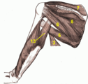 Muscles on the dorsum of thetriceps brachii muscle
 -
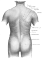 Surface anatomy of the back. (Label for Teres major at upper right.)
 -
 Left humerus. Anterior view.
 -
 Teres major muscle
 -
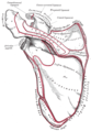 Left scapula. Posterior surface.
 -
 Teres major muscle


See also
- Accessory muscles of the scapula
References
![]() This article incorporates text in the public domain from page 442 of the 20th edition of Gray's Anatomy (1918)
This article incorporates text in the public domain from page 442 of the 20th edition of Gray's Anatomy (1918)
- PMID 23931789, retrieved November 2, 2020
- ISBN 978-0-12-802653-3, retrieved November 2, 2020
- ^ ISBN 978-0-7506-7332-7, retrieved November 2, 2020
- S2CID 3872258.
External links
- Anatomy figure: 03:03-06 at Human Anatomy Online, SUNY Downstate Medical Center
- PTCentral