
Medulla oblongata
This article needs additional citations for verification. (October 2010) |
| Medulla oblongata | |
|---|---|
Brain stem | |
| Identifiers | |
| Latin | medulla oblongata, myelencephalon |
| MeSH | D008526 |
| NeuroNames | 698 |
| NeuroLex ID | birnlex_957 |
| TA98 | A14.1.03.003 |
| TA2 | 5983 |
| FMA | 62004 |
| Anatomical terms of neuroanatomy] | |
The medulla oblongata or simply medulla is a long stem-like structure which makes up the lower part of the
During embryonic development, the medulla oblongata develops from the
The bulb is an archaic term for the medulla oblongata..
Anatomy


The medulla can be thought of as being in two parts:
- an upper open part or superior part where the dorsal surface of the medulla is formed by the fourth ventricle.
- a lower closed part or inferior part where the fourth ventricle has narrowed at the central canal.
External surfaces
The
The region between the anterolateral and posterolateral sulcus in the upper part of the medulla is marked by a pair of swellings known as olivary bodies (also called olives). They are caused by the largest nuclei of the olivary bodies, the inferior olivary nuclei.
The posterior part of the medulla between the
Just above the tubercles, the posterior aspect of the medulla is occupied by a triangular fossa, which forms the lower part of the floor of the fourth ventricle. The fossa is bounded on either side by the inferior cerebellar peduncle, which connects the medulla to the cerebellum.
The lower part of the medulla, immediately lateral to the cuneate fasciculus, is marked by another longitudinal elevation known as the
Blood supply
Blood to the medulla is supplied by a number of
- Anterior spinal artery: This supplies the whole medial part of the medulla oblongata.
- Posterior inferior cerebellar artery: This is a major branch of the vertebral artery, and supplies the posterolateral part of the medulla, where the main sensory tracts run and synapse. It also supplies part of the cerebellum.
- Direct branches of the vertebral artery: The vertebral artery supplies an area between the anterior spinal and posterior inferior cerebellar arteries, including the solitary nucleus and other sensory nuclei and fibers.
- cuneate nucleus.
Development
The medulla oblongata forms in fetal development from the myelencephalon. The final differentiation of the medulla is seen at week 20 gestation.[4][full citation needed]
Neuroblasts from the alar plate of the neural tube at this level will produce the sensory nuclei of the medulla. The basal plate neuroblasts will give rise to the motor nuclei.
- Alar plate neuroblasts give rise to:
- The special visceral afferentcolumn.
- The spinal trigeminal nerve nuclei which contains the general somatic afferent column.
- The special somatic afferentcolumn.
- The inferior olivary nucleus, which relays to the cerebellum.
- The cuneate nuclei.
- The
- Basal plate neuroblasts give rise to:
- The general somatic efferent fibers.
- The special visceral efferent.
- The general visceral efferent fibers.
- The
Function
The medulla oblongata connects the higher levels of the
- The dorsal respiratory group are neurons involved in this regulation. The pre-Bötzinger complexis a cluster of interneurons involved in the respiratory function of the medulla.
- Cardiovascular center – sympathetic, parasympathetic nervous system
- Vasomotor center – baroreceptors
- masseter reflex can be termed bulbar reflexes.[5]
Clinical significance
A
Lateral medullary syndrome can be caused by the blockage of either the posterior inferior cerebellar artery or of the vertebral arteries.
Progressive bulbar palsy (PBP) is a disease that attacks the nerves supplying the bulbar muscles. Infantile progressive bulbar palsy is progressive bulbar palsy in children.
Other animals
Both lampreys and hagfish possess a fully developed medulla oblongata.
Additional images
-
 Lobes
Lobes -
 Cross section of the medulla (in red) and surrounding tissues.
 -
 Anteroinferior viewof the medulla oblongata and pons.
 -
 Base of brain.
 -
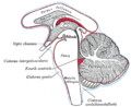 Diagram showing the positions of the three principal subarachnoid cisternæ.
 -
 Medulla oblongata
 -
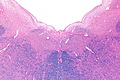 H&E-LFB stain.


References
![]() This article incorporates text in the public domain from page 767 of the 20th edition of Gray's Anatomy (1918)
This article incorporates text in the public domain from page 767 of the 20th edition of Gray's Anatomy (1918)
- ^ ISBN 978-0-323-10027-4, retrieved 2020-11-15
- ^ ISBN 978-1-4160-5893-9, retrieved 2020-11-15
- ^ Purves, Dale (2001). Neuroscience. 2nd edition. Sinauer Associates.
- ^ Carlson, Neil R. Foundations of Behavioral Neuroscience.63-65
- S2CID 45624479.)
{{cite journal}}: CS1 maint: multiple names: authors list (link - S2CID 27654584.
- ^ Haycock, Being and Perceiving
- Haycock DE (2011). Being and Perceiving. Manupod Press. ISBN 978-0-9569621-0-2.