
Upper extremity of femur
| Upper extremity of femur | |
|---|---|
| Details | |
| Identifiers | |
| Latin | Extremitas proximalis ossis femoris |
| FMA | 32841 |
| Anatomical terms of bone] | |
The upper extremity, proximal extremity or superior epiphysis of the femur is the part of the
- Femoral head including the fovea
- Femur neck
- Greater trochanter
- Lesser trochanter
- Intertrochanteric line
- Intertrochanteric crest
- Trochanteric fossa
- Linea quadrata
- Quadrate tubercle
The
In the transition area between the head and neck is quite rough due to attachment of muscles and the
A slight ridge is sometimes seen commencing about the middle of the intertrochanteric crest, and reaching vertically downward for about 5 cm. along the back part of the body: it is called the
About the junction of the upper one-third and lower two-thirds on the intertrochanteric crest is the quadrate tubercle located. The size of the tubercle varies and it is not always located on the intertrochanteric crest and that also adjacent areas can be part of the quadrate tubercle, such as the posterior surfare of the greater trochanter or the neck of the femur. In a small anatomical study it was shown that the epiphysial line passes directly through the quadrate tubercle.[2]
Additional images
-
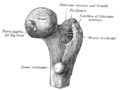 Upper extremity of right femur viewed from behind and above, showingneck, and the greater and lesser trochanter
 -
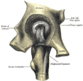 Left hip-joint, opened by removing the floor of the acetabulum from within the pelvis.
 -
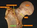 Superior epiphysis - anterior view
 -
 Superior epiphysis - posterior view


References
- ^ ISBN 978-87-628-0307-7.
- PMID 17104699.