
First metatarsal bone
| First metatarsal bone | |
|---|---|
 The first metatarsal. (Left.) | |
 Bones of the right foot. Dorsal surface. The first metatarsal bone is shown in yellow farthest to the left | |
| Details | |
| Identifiers | |
| Latin | os metatarsale I |
| TA2 | 1500 |
| FMA | 24502 |
| Anatomical terms of bone | |
The first metatarsal bone is the
Like the four other metatarsals, it can be divided into three parts: base, body and head. The base is the part closest to the ankle and the head is closest to the big toe. The narrowed part in the middle is referred to as the body of the bone. The bone is somewhat flattened, giving it two sides: the plantar (towards the sole of the foot) and the dorsal side (the area facing upwards while standing).[1]
The base presents, as a rule, no
The first metatarsal articulates (forms
The body of the bone is strong, and of well-marked prismoid form.
The head is large; on its plantar surface are two grooved facets on which the sesamoid bones glide; the facets are separated by a smooth elevation.
Muscle attachments
 |
 |
Three muscles attach to the first metatarsal bone: the tibialis anterior, fibularis longus and first dorsal interosseus.[3]
The tibialis anterior inserts at the basis of the bone, while the fibularis longus inserts at the tuberosity. The lateral part of the first dorsal interosseus muscle originates from the medial side of the bone. Its function is to spread the toes.[4]
| Muscle | Direction | Attachment[3] |
|---|---|---|
| Tibialis anterior | Insertion | Basis of first metatarsal |
| Fibularis longus | Insertion | Tuberosity of first metatarsal |
| Dorsal interossei I | Origin | Lateral part of first metatarsal |
Additional images
-
 Sesamoid bones at the distal end of the first metatarsal.
 -
 First metatarsal bone. Deep dissection.
 -
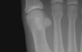
![It is common in children to have a pseudo-epiphysis of the first metatarsal.[5]](//upload.wikimedia.org/wikipedia/commons/thumb/4/4f/Metatarsal_pseudo-epiphysis.jpg/94px-Metatarsal_pseudo-epiphysis.jpg) It is common in children to have apseudo-epiphysis of the first metatarsal.[5]
It is common in children to have apseudo-epiphysis of the first metatarsal.[5]


![It is common in children to have a pseudo-epiphysis of the first metatarsal.[5]](/File:Metatarsal_pseudo-epiphysis.jpg)
References
![]() This article incorporates text in the public domain from page 272 of the 20th edition of Gray's Anatomy (1918)
This article incorporates text in the public domain from page 272 of the 20th edition of Gray's Anatomy (1918)
- ^ ISBN 978-87-628-0307-7.
- ^ Platzer 2004, p. 218
- ^ ISBN 978-87-628-0307-7.
- ISBN 978-87-628-0307-7.
- PMID 2681682.