
Anatomy of the cerebellum
| Cerebellum | |
|---|---|
inferior | |
| Identifiers | |
| NeuroLex ID | birnlex_1489 |
| TA98 | A14.1.07.001 |
| TA2 | 5788 |
| Anatomical terms of neuroanatomy] | |
The anatomy of the cerebellum can be viewed at three levels. At the level of
Gross anatomy
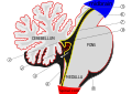
The cerebellum is located at the base of the brain, with the large mass of the
Because of its large number of tiny granule cells, the cerebellum contains more neurons than the rest of the brain put together, but it only takes up 10% of total brain volume.[3] The cerebellum receives nearly 200 million input fibers; in contrast, the optic nerve is composed of a mere one million fibers.
The unusual surface appearance of the cerebellum conceals the fact that the bulk of the structure is made up of a very tightly folded layer of gray matter, the
The cerebellum can be divided according to three different criteria: gross anatomical, phylogenetical, and functional.
Gross anatomical divisions
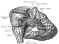
On gross inspection, three lobes can be distinguished in the cerebellum: the flocculonodular lobe, the anterior lobe (rostral to the "primary fissure"), and the posterior lobe (dorsal to the "primary fissure"). The latter two can be further divided in a midline cerebellar vermis and lateral cerebellar hemispheres.
Tonsil . H: Anterior lobe. I: Posterior lobe. |
 |
Phylogenetic and functional divisions
The cerebellum can also be divided in three parts based on both phylogenetic criteria (the evolutionary age of each part) and on functional criteria (the incoming and outgoing connections each part has and the role played in normal cerebellar function). From the phylogenetically oldest to the newest, the three parts are:
| Functional denomination (phylogenetic denomination) | Anatomical parts | Role |
| Vestibulocerebellum (Archicerebellum) | Flocculonodular lobe (and immediately adjacent vermis) | The vestibulocerebellum regulates balance and eye movements. It receives superior colliculi and from the visual cortex (the latter via the pontine nuclei, forming a cortico-ponto-cerebellar pathway). Lesions of the vestibulocerebellum cause disturbances of balance and gait. There is another small region, known as the biventer lobule .
|
| Spinocerebellum (Paleocerebellum) | Vermis and intermediate parts of the hemispheres ("paravermis") |
The spinocerebellum regulates body and limb movements. It receives proprioception input from the dorsal columns of the spinal cord (including the spinocerebellar tract) and the trigeminal nerve, as well as from visual and auditory systems. It sends fibres to deep cerebellar nuclei (including the fastigial nucleus) which in turn project to both the cerebral cortex (via midbrain and thalamus) and the brain stem (via reticular formation in the pons, and vestibular nuclei in the medulla oblongata ), thus providing modulation of descending motor systems. The spinocerebellum contains sensory maps as it receives data on the position of various body parts in space: in particular, the vermis receives fibres from the trunk and proximal portions of limbs, while the intermediate parts of the hemispheres receive fibres from the distal portions of limbs. The spinocerebellum is able to elaborate proprioceptive input in order to anticipate the future position of a body part during the course of a movement, in a "feed forward" manner.
|
| Cerebrocerebellum (Neocerebellum, Pontocerebellum) | Lateral parts of the hemispheres | The neocerebellum is involved in planning movement and evaluates sensory information for action. It receives input exclusively from the cerebral cortex (especially the primary motor area of the cerebral cortex) and to the red nucleus (in turn connected to the inferior olivary nucleus, which links back to the cerebellar hemispheres). The neocerebellum is involved in planning movement that is about to occur[7] and has purely cognitive functions as well.
|
Much of what is understood about the functions of the cerebellum stems from careful documentation of the effects of focal lesions in human patients who have suffered from injury or disease or through animal lesion research.
Cellular anatomy
As explained in more detail in the Function section, the cerebellum differs from most other brain areas in that the flow of neural signals through it is almost entirely unidirectional: there are virtually no backward connections between its neuronal elements. Thus the most logical way to describe the cellular structure is to begin with the inputs and follow the sequence of connections through to the outputs.
Deep nuclei
The four
Cortical layers
The
There are three layers to the cerebellar cortex; from outer to inner layer, these are the molecular, Purkinje, and granular layers. The function of the cerebellar cortex is essentially to modulate information flowing through the deep nuclei. The microcircuitry of the cerebellum is schematized in Figure 5. Mossy and climbing fibers carry sensorimotor information into the deep nuclei, which in turn pass it on to various premotor areas, thus regulating the gain and timing of motor actions. Mossy and climbing fibers also feed this information into the cerebellar cortex, which performs various computations, resulting in the regulation of Purkinje cell firing. Purkinje neurons feed back into the deep nuclei via a potent inhibitory synapse. This synapse regulates the extent to which mossy and climbing fibers activate the deep nuclei, and thus control the ultimate effect of the cerebellum on motor function. The synaptic strength of almost every synapse in the cerebellar cortex has been shown to undergo synaptic plasticity. This allows the circuitry of the cerebellar cortex to continuously adjust and fine-tune the output of the cerebellum, forming the basis of some types of motor learning and coordination. Each layer in the cerebellar cortex contains the various cell types that comprise this circuitry.
Molecular layer
This outermost layer of the cerebellar cortex contains two types of inhibitory
Purkinje layer
The middle layer contains only one type of cell body—that of the large Purkinje cell. Purkinje cells are the primary integrative neurons of the cerebellar cortex and provide its sole output. Purkinje cell dendrites are large arbors with hundreds of spiny branches reaching up into the molecular layer (Fig. 6). These dendritic arbors are flat—nearly all of them lie in planes—with neighboring Purkinje arbors in parallel planes. Each parallel fiber from the granule cells runs orthogonally through these arbors, like a wire passing through many layers. Purkinje neurons are GABAergic—meaning they have inhibitory synapses—with the neurons of the deep cerebellar and vestibular nuclei in the brainstem. Each Purkinje cell receives excitatory input from 100,000 to 200,000 parallel fibers. Parallel fibers are said to be responsible for the simple (all or nothing, amplitude invariant) spiking of the Purkinje cell.
Purkinje cells also receive input from the inferior olivary nucleus via climbing fibers. A good mnemonic for this interaction is the phrase "climb the other olive tree", given that climbing fibers originate from the contralateral inferior olive. In striking contrast to the 100,000-plus inputs from parallel fibers, each Purkinje cell receives input from exactly one climbing fiber; but this single fiber "climbs" the dendrites of the Purkinje cell, winding around them and making a large number of synapses as it goes. The net input is so strong that a single action potential from a climbing fiber is capable of producing a "complex spike" in the Purkinje cell: a burst of several spikes in a row, with diminishing amplitude,[9] followed by a pause during which simple spikes are suppressed.
Just underneath the Purkinje layer are the Lugaro cells whose very long dendrites travel along the boundary between the Purkinje and the granular layers.
Granular layer
The innermost layer contains the cell bodies of three types of cells: the numerous and tiny
Relationship with cerebral cortex
The local field potentials of the neocortex and cerebellum oscillate coherently at (6–40 Hz) in awake behaving animals.[11] These appear to be under the control of output from the cerebral cortex.[12] This output would be mediated by a pathway from layer 5/6 neurons in the neocortex through that project either to the pons or the inferior olive. If through the pons this would go to mossy fibers that synapse with granule and Golgi neurons with the granule cells then targeting Purkinje neurons via their excitatory parallel fibers. If the inferior olive it would go via excitatory climbing fiber inputs to Purkinje neurons.[12] These return this output back to the cerebral cortex through the ventrolateral thalamus completing the loop.
The corticopontocerebellar pathway is the largest pathway associated with the cerebellum. Arising in the cerebral cortex these fibers first terminate ipsilaterally in the pontine nuclei. Then the fibers decussate and form the middle cerebellar peduncle, terminating in the cerebellar cortex as mossy fibers. This pathway transmits signals that inform the cerebellum about the movement in progress and the upcoming movement. This helps the continuous adjustment of motor activity.[13]
The initiation of the movement is relayed to cerebellum via the corticoreticulocerebellar pathway. Those synapse ipsilaterally in the reticular formation, then via the inferior and middle peduncles into the cerebellar vermis.[13]
The
The cerebellum send its projections back to the cerebral cortex via the Cerebellothalamic tract.
The cerebellar lateral expansion, or the neocerebellum, may be associated with cognitive functions, and it is anatomically linked with the lateral prefrontal cortex. It shows greatest activity during speech, with a one-sided predominance consistent with a possible linkage (via the thalamus) with the motor speech area.[14]
When lesions occur in the association areas linked to the cerebellum by corticopontocerebellar fibres, the cognitive affective syndrome may occur. This results in cognitive defects in the form of diminished reasoning power, inattention, grammatical errors in speech, poor spatial sense, and patchy memory loss.[14]
Blood supply


Three arteries supply blood to the cerebellum (Fig. 7): the superior cerebellar artery (SCA), anterior inferior cerebellar artery (AICA), and posterior inferior cerebellar artery (PICA).
The SCA branches off the lateral portion of the basilar artery, just inferior to its bifurcation into the posterior cerebral artery. Here, it wraps posteriorly around the pons (to which it also supplies blood) before reaching the cerebellum. The SCA supplies blood to most of the cerebellar cortex, the cerebellar nuclei, and the superior cerebellar peduncles.[15]
The AICA branches off the lateral portion of the basilar artery, just superior to the junction of the vertebral arteries. From its origin, it branches along the inferior portion of the pons at the
The PICA branches off the lateral portion of the vertebral arteries just inferior to their junction with the basilar artery. Before reaching the inferior surface of the cerebellum, the PICA sends branches into the medulla, supplying blood to several
Variations among vertebrates
There is considerable variation in the size and shape of the cerebellum in different vertebrate species. It is generally largest in
In amphibians, lampreys, and hagfish the cerebellum is little developed; in the latter two groups it is barely distinguishable from the brain-stem. Although the spinocerebellum is present in these groups, the primary structures are small paired nuclei corresponding to the vestibulocerebellum.[16]
Peduncles
The cerebellum follows the general groups-of-three pattern found in anatomy,[17] with three major input and output cerebellar peduncles (fiber bundles). These are the superior (brachium conjunctivum), middle (brachium pontis), and inferior (restiform and juxtarestiform bodies) cerebellar peduncles.
| Peduncle | Description |
Superior |
While there are some afferent fibers from the cerebellothalamocortical (cerebellum > thalamus > premotor cortex) pathways are two major pathways that pass through this peduncle and are important in motor planning .
|
Middle |
This is composed entirely of afferent fibers originating within the neocortex and make the middle cerebellar peduncle the largest of the three cerebellar peduncles.
|
| Inferior | This carries many types of input and output fibers that are mainly concerned with integrating Purkinje cells out to the vestibular nuclei in the dorsal brainstem located at the junction between the pons and medulla .
|
There are three sources of input to the cerebellum, in two categories consisting of mossy and climbing fibers, respectively. Mossy fibers can originate from the pontine nuclei, which are clusters of neurons located in the pons that carry information from the contralateral cerebral cortex. They may also arise within the spinocerebellar tract whose origin is located in the
Development
During the early stages of
Two primary regions are thought to give rise to the neurons that make up the cerebellum. The first region is the
Additional images
-
 Dissection showing the projection fibers of the cerebellum
 -
 Scheme of roof of fourth ventricle. The arrow is in the foramen of Majendie.
 -
 Human brain midsagittal view
 -
 Anterior view of the human cerebellum, with numbers indicating salient landmarks


References
- ^ a b Knierim, James. "Chapter 5: Cerebellum". Neuroscience Online: An Electronic Textbook for the Neurosciences.
- S2CID 5387374.
- ^ The Brain From Top To Bottom
- PMID 38453991.
- PMID 18222655.
- PMID 12902393.
- ISBN 0-683-30460-7.
- ^ Harting, J.K. "The Global Cerebellum '97". University of Wisconsin Medical School.
- PMID 18650337.
- PMID 9310423.
- PMID 16424458.
- ^ PMID 19692605.
- ^ ISBN 9781405103404.
- ^ a b c Mtui, Estomih; Gruener, Gregory; Dockery, Peter (2016). Fitzgerald's Clinical Neuroanatomy and Neuroscience (7th ed.). Elsevier. pp. 243–252.
- ^ Gray, Henry; Lewis, Warren Harmon (1918). Anatomy of the human body (20th ed.). Philadelphia: Lea & Febiger.
- ^ ISBN 0-03-910284-X.
- ^ "List of Three's". www.meddean.luc.edu.
- S2CID 33485509.
- PMID 2723742.