
Superior cerebellar peduncle
| Superior cerebellar peduncle | |
|---|---|
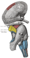
vermis with the hemisphere. (Superior peduncle labeled at upper right.) | |
 Dissection showing the projection fibers of the cerebellum. (Superior peduncle labeled at center top.) | |
| Details | |
| Identifiers | |
| Latin | pedunculus cerebellaris superior |
| NeuroNames | 833 |
| NeuroLex ID | birnlex_1711 |
| TA98 | A14.1.05.006 A14.1.07.417 A14.1.08.678 A14.1.06.009 A14.1.06.216 |
| TA2 | 5846 |
| FMA | 72495 |
| Anatomical terms of neuroanatomy] | |
In the
ventral spinocerebellar tract. Other afferent tracts are the trigeminothalamic fibers, tectocerebellar fibers, and noradrenergic fibers from the locus coeruleus. The superior peduncle emerges from the upper and medial parts of the white matter of each hemisphere[citation needed
] and is placed under cover of the upper part of the cerebellum.
Structure
Superior cerebellar peduncles are connected together by the
inferior colliculi
, under which they disappear.
Below, they form the upper lateral boundaries of the fourth ventricle, but as they ascend they converge on the dorsal aspect of the ventricle and thus assist in forming its roof.
Decussation

The decussation of superior cerebellar peduncle is the crossing of fibers of the superior cerebellar peduncle across the midline, and is located at the level of the
contralateral red nucleus to eventually become the rubrospinal tract. It is also known as horseshoe-shaped commissure of Wernekinck.[1]
It is important as an anatomical landmark, as lesions above it cause contralateral cerebellar signs, while lesions below it cause ipsilateral cerebellar signs.
Additional images
-
 Scheme showing the connections of the several parts of the brain.
 -
 Superficial dissection of brain-stem. Lateral view.
 -
 Axial section of the pons, at its dorsal (superior) part.
 -
 Sagittal section through right cerebellar hemisphere. The right olive has also been cut sagittally.
 -
 Dissection showing the course of the cerebrospinal fibers.
 -
 Upper part of medulla spinalis and hind- and mid-brains; posterior aspect, exposed in situ.


References
![]() This article incorporates text in the public domain from page 792 of the 20th edition of Gray's Anatomy (1918)
This article incorporates text in the public domain from page 792 of the 20th edition of Gray's Anatomy (1918)
- S2CID 15717777.
External links
- Atlas image: n2a7p6 at the University of Michigan Health System
- Atlas image: n2a7p4 at the University of Michigan Health System
- https://web.archive.org/web/20080518232033/http://isc.temple.edu/neuroanatomy/lab/atlas/micn/
- http://www.neuroanatomy.wisc.edu/Bs97/TEXT/P18/sum.htm
- NIF Search - Decussation of superior cerebellar peduncle via the Neuroscience Information Framework