
Spinocerebellar tract
| Spinocerebellar tract | |
|---|---|
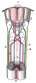
 Spinocerebellar tracts are labeled in blue at right. | |
| Details | |
| Identifiers | |
| Latin | tractus spinocerebellaris |
| MeSH | D020824 |
| NeuroNames | 1978 |
| Anatomical terms of neuroanatomy | |
The spinocerebellar tract is a
Origins of proprioceptive information
Proprioceptive information is obtained by Golgi tendon organs and muscle spindles.
- Golgi tendon organs consist of a fibrous capsule enclosing tendon fascicles and bare nerve endings that respond to tension in the tendon by causing action potentials in type Ib afferents. These fibers are relatively large, myelinated, and quickly conducting.
- nuclear bag fibers and nuclear chain fibers) and type II afferents(solely from nuclear chain fibers).
All of these neurons are sensory (first order, or primary) and have their cell bodies in the dorsal root ganglia. They pass through Rexed laminae layers I-VI of the posterior grey column (dorsal horn) to form synapses with second order or secondary neurons in layer VII just beneath the dorsal horn.
Subdivisions of the tract
The tract is divided into:[1] [dubious ]
| Division | Peripheral Process of First Order the Neuron | Region of Innervation |
|---|---|---|
dorsal (posterior) spinocerebellar tract |
from Golgi tendon organs |
ipsilateral caudal aspect of the body and legs |
ventral (anterior) spinocerebellar tract |
from Golgi tendon organs |
ipsilateral caudal aspect of the body and legs |
Cuneocerebellar tract |
from Golgi tendon organs |
ipsilateral arm |
Rostral spinocerebellar tract |
from Golgi tendon organs |
ipsilateral arm |
Dorsal spinocerebellar tract
The dorsal spinocerebellar tract (posterior spinocerebellar tract, Flechsig's fasciculus, Flechsig's tract) conveys proprioceptive information from proprioceptors in the skeletal muscles and joints to the cerebellum.[2]
It is part of the
This tract involves two neurons and ends up on the same side of the body.
The terms Flechsig's fasciculus and Flechsig's tract are named after German
Ventral spinocerebellar tract
The ventral spinocerebellar tract (or anterior spinocerebellar tract) conveys
It is part of the
The ventral tract (under L2/L3) gets its proprioceptive/fine touch/vibration information from a first order neuron, with its cell body in a dorsal ganglion. The axon runs via the fila radicularia to the dorsal horn of the grey matter. There it makes a synapse with the dendrites of two neurons: they send their axons bilaterally to the ventral border of the lateral funiculi. The fibers of the ventral spinocerebellar tract then enters the cerebellum via the
Originates from ventral horn at lumbosacral spinal levels. Axons first cross midline in the spinal cord and run in the ventral border of the lateral funiculi. These axons ascend to the pons where they join the superior cerebellar peduncle to enter the cerebellum. Once in the deep white matter of the cerebellum, the axons recross the midline, give off collaterals to the globose and
Comparison with dorsal spinocerebellar tract
When the dorsal roots are cut in a cat performing a step cycle, peripheral excitation is lost, and the dorsal spinocerebellar tract has no activity; the ventral spinocerebellar tract continues to show activity. This suggests that the dorsal spinocerebellar tract carries sensory information to the spinocerebellum through the inferior cerebellar peduncle during movement (since the inferior peduncle is known to contain fibres from the dorsal tract), and that the ventral spinocerebellar tract carries internally generated motor information about the movement through the superior cerebellar peduncle.[3]
Posterior external arcuate fibers
The posterior external arcuate fibers (dorsal external arcuate fibers or cuneocerebellar tract)
The posterior external arcuate fibers carry proprioceptive information from the upper limbs and neck. It is an analogue to the
It is uncertain whether fibers are continued directly from the gracile and cuneate fasciculi into the inferior peduncle.
Rostral spinocerebellar tract
The rostral spinocerebellar tract is a tract which transmits information from the
Pathway for dorsal and spinocuneocerebellar tracts
The sensory neurons synapse in an area known as
This is a column of relay neuron cell bodies within the medial gray matter within the spinal cord in layer VII (just beneath the dorsal horn), specifically between T1-L3. These neurons then send axons up the spinal cord, and project ipsilaterally to medial zones of the cerebellum through the inferior cerebellar peduncle.
Below L3, relevant neurons pass into the
The neurons in the accessory cuneate nucleus have axons leading to the ipsilateral cerebellum via the inferior cerebellar peduncle.
Pathway for ventral and rostral spinocerebellar tracts
Some neurons of the ventral spinocerebellar tract instead form synapses with neurons in layer VII of L4-S3. Most of these fibers cross over to the contralateral lateral funiculus via the anterior white commissure and through the superior cerebellar peduncle. The fibers then often cross over again within the cerebellum to end on the ipsilateral side. For this reason the tract is sometimes termed the "double-crosser."
The Rostral Tract synapses at the dorsal horn lamina (intermediate gray zone) of the spinal cord and ascends ipsilaterally to the cerebellum through the inferior cerebellar peduncle
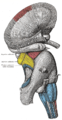
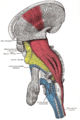
Additional images
-
 Decussation of pyramids.
 -
 Superficial dissection of brain-stem. Lateral view.
 -
 Deep dissection of brain-stem. Lateral view.
 -
 Dissection of brain-stem. Lateral view.
 -
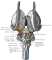 Dissection of brain-stem. Dorsal view.


References
- ^ Siegel, Allan, and Hreday N. Sapru. Essential Neuroscience. 2nd. Lippincott, 2011. 146-149.
- ISBN 970-10-5504-7
- ISBN 0-8385-7701-6.
- ISBN 978-1-58890-572-7.
- S2CID 23836263.
- ISBN 978-0-7817-2829-4.
- ^ "Archived copy". Archived from the original on 2008-04-30. Retrieved 2019-10-02.
{{cite web}}: CS1 maint: archived copy as title (link) - ISBN 0-86577-710-1.
- ^ a b "Rostral spinocerebellar tract". The Neuroscience Lexicon. Retrieved 19 May 2013.
Further reading
- OSCARSSON, O.; UDDENBERG, N. (1 May 1965). "Properties of Afferent Connections to the Rostral Spinocerebellar Tract in the Cat". Acta Physiologica Scandinavica. 64 (1–2): 143–153. PMID 14347272.
External links
- hier-804 at NeuroNames
- hier-805 at NeuroNames
- hier-793 at NeuroNames - dorsal external arcuate fibers
- hier-800 at NeuroNames - cuneocerebellar tract
- Anatomyatlases Plate17327
- NIF Search - Anterior Spinocerebellar Tract via the Neuroscience Information Framework