
Body of sphenoid bone
| Body of sphenoid bone | |
|---|---|
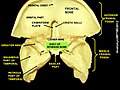
 Figure 2: Sphenoid bone, anterior and inferior surfaces. | |
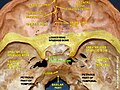
 Figure 3: Sphenoid bone, upper and posterior surfaces. | |
| Details | |
| Identifiers | |
| Latin | corpus ossis sphenoidalis |
| TA98 | A02.1.05.002 |
| TA2 | 585 |
| FMA | 52867 |
| Anatomical terms of bone | |
The body of the
Superior surface
The superior surface of the body [Fig. 1] presents in front a prominent spine, the ethmoidal spine, for articulation with the cribriform plate of the ethmoid bone; behind this is a smooth surface slightly raised in the middle line, and grooved on either side for the olfactory lobes of the brain.
This surface is bounded behind by a ridge, which forms the anterior border of a narrow, transverse groove, the
Behind the chiasmatic groove is an elevation, the
The anterior boundary of the sella turcica is completed by two small eminences, one on either side, called the middle clinoid processes, while the posterior boundary is formed by a square-shaped plate of bone, the dorsum sellae, ending at its superior angles in two tubercles, the posterior clinoid processes, the size and form of which vary considerably in different individuals.
The posterior clinoid processes deepen the sella turcica, and give attachment to the
On either side of the dorsum sellae is a notch for the passage of the
Behind the dorsum sellae is a shallow depression, the clivus, which slopes obliquely backward, and is continuous with the groove on the basilar portion of the occipital bone; it supports the upper part of the pons.
Lateral surfaces
The lateral surfaces of the body are united with the
Above the attachment of each greater wing is a broad groove, curved something like the italic letter f; it lodges the internal carotid artery and the cavernous sinus, and is named the carotid sulcus.
Along the posterior part of the lateral margin of this groove, in the angle between the body and greater wing, is a ridge of bone, called the sphenoidal lingula.
Posterior surfaces
The posterior surface, quadrilateral in form [Fig. 3], is joined, during infancy and adolescence, to the basilar part of the occipital bone by a plate of cartilage.
Between the eighteenth and twenty-fifth years this becomes ossified, ossification commencing above and extending downward.
Anterior surface
The anterior surface of the body [Fig. 2] presents, in the middle line, a vertical crest, the sphenoidal crest, which articulates with the perpendicular plate of the
On either side of the crest is an irregular opening leading into the corresponding
These sinuses are two large, irregular cavities hollowed out of the interior of the body of the bone, and separated from one another by a bony septum, which is commonly bent to one or the other side.
They vary considerably in form and size, are seldom symmetrical, and are often partially subdivided by irregular bony laminae.
Occasionally, they extend into the basilar part of the occipital bone nearly as far as the foramen magnum. They begin to be developed before birth, and are of a considerable size by the age of six.
They are partially closed, in front and below, by two thin, curved plates of bone, the sphenoidal conchae, leaving in the articulated skull a round opening at the upper part of each sinus by which it communicates with the upper and back part of the nasal cavity and occasionally with the posterior ethmoidal air cells.
The lateral margin of the anterior surface is serrated, and articulates with the
Inferior surface
The inferior surface presents, in the middle line, a triangular spine, the sphenoidal rostrum, which is continuous with the sphenoidal crest on the anterior surface, and is received in a deep fissure between the alæ of the vomer.
On either side of the rostrum is a projecting lamina, the vaginal process, directed medialward from the base of the
Additional images
-
 Body of sphenoid bone
 -
 Body of sphenoid bone
 -
 Body of sphenoid bone


References
- ISBN 9781455710676.