
Facial canal
| Facial canal | |
|---|---|
skeletal | |
| Nerve | facial nerve (CN VII) |
| Identifiers | |
| Latin | canalis nervi facialis, canalis facialis |
| TA98 | A02.1.06.009 |
| TA2 | 688 |
| FMA | 54952 |
| Anatomical terminology] | |
The facial canal (also known as the Fallopian canal) is a Z-shaped canal in the
Anatomy
The facial canal gives passage to the
The facial nerve gives rise to three nerves while passing through the canal: the
Structure
Horizontal part
The proximal portion of the facial canal is termed the horizontal part. It commences at the introitus of facial canal at the distal end of the internal auditory meatus. The horizontal part is further subdivided into two crura: the proximal/medial[4] anteriolaterally[5] directed medial crus (or labyrinthine segment[5]), and the distal/lateral[4] posteriolaterally[5] directed lateral crus (or tympanic segment[5]); the two crura meet at a sharp angle at the genu of facial canal (geniculum canalis facialis[6]) where the geniculate ganglion is situated (at the genu, the greater petrosal nerve leaves the facial canal through the hiatus of the facial canal).[4]
Descending part
The lateral crus of horizontal part ends by turning sharply inferior-ward, commencing the distal-most descending part (or mastoid segment
Relations
The labyrinthine segment is situated superior to cochlea.[5]
The canal traverses the
Clinical significance
The facial canal may be interrupted in some people. This may lead to the facial nerve being split into 2 or 3 fibres, or it may be poorly formed or congenitally absent on one side.[2]
History
The facial canal was first described by Gabriele Falloppio.[10] This is why it may also be known as the Fallopian canal.[10]
Gallery
-
 Lateral head anatomy detail. Facial nerve dissection.
 -
 Tympanic cavity. Facial canal. Internal carotid artery.
 -
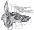 Coronal section of right temporal bone. Prominence of the facial canal labeled at top, fourth from the left.


See also
- Facial nerve
- Prominence of the facial canal
- Hiatus of the facial canal
References
- PMID 1762775.
- ^ S2CID 25764734.
- ISBN 978-1-4963-4721-3.
- ^ a b c "horizontal part of facial canal". TheFreeDictionary.com. Retrieved 2023-07-29.
- ^ S2CID 205411993.
- ^ "genu of facial canal". TheFreeDictionary.com. Retrieved 2023-07-29.
- ^ "posterior canaliculus of chorda tympani". TheFreeDictionary.com. Retrieved 2023-07-29.
- ^ "descending part of facial canal". TheFreeDictionary.com. Retrieved 2023-07-29.
- OCLC 1201341621.)
{{cite book}}: CS1 maint: location missing publisher (link - ^ S2CID 12712839.