
Occipital bone
| Occipital bone | |
|---|---|
 Position of occipital bone | |
 Animation of the occipital bone | |
| Details | |
| Articulations | The two parietals, the two temporals, the sphenoid, and the atlas |
| Identifiers | |
| Latin | os occipitale |
| MeSH | D009777 |
| TA98 | A02.1.04.001 |
| TA2 | 552 |
| FMA | 52735 |
| Anatomical terms of bone | |
The occipital bone (
Like the other cranial bones, it is classed as a flat bone. Due to its many attachments and features, the occipital bone is described in terms of separate parts. From its front to the back is the basilar part, also called the basioccipital, at the sides of the foramen magnum are the lateral parts, also called the exoccipitals, and the back is named as the squamous part. The basilar part is a thick, somewhat quadrilateral piece in front of the foramen magnum and directed towards the pharynx. The squamous part is the curved, expanded plate behind the foramen magnum and is the largest part of the occipital bone.
Due to its embryonic derivation from paraxial mesoderm (as opposed to neural crest, from which many other craniofacial bones are derived), it has been posited that "the occipital bone as a whole could be considered as a giant vertebra enlarged to support the brain."[1]
Structure
The occipital bone, like the other seven
Outer surface

Near the middle of the outer surface of the squamous part of the occipital (the largest part) there is a prominence – the external occipital protuberance. The highest point of this is called the inion.
From the inion, along the midline of the squamous part until the foramen magnum, runs a ridge – the external occipital crest (also called the medial nuchal line) and this gives attachment to the nuchal ligament.
Running across the outside of the occipital bone are three curved lines and one line (the medial line) that runs down to the foramen magnum. These are known as the
Inner surface

The inner surface of the occipital bone forms the base of the posterior cranial fossa. The foramen magnum is a large hole situated in the middle, with the clivus, a smooth part of the occipital bone travelling upwards in front of it. The median internal occipital crest travels behind it to the internal occipital protuberance, and serves as a point of attachment to the falx cerebri.
To the sides of the
The inner surface of the occipital bone is marked by dividing lines as shallow ridges, that form four fossae or depressions. The lines are called the cruciform (cross-shaped) eminence.
At the midpoint where the lines intersect a raised part is formed called the internal occipital protuberance. From each side of this eminence runs a groove for the transverse sinuses.
There are two midline
Foramen magnum
The foramen magnum (
Through the foramen passes the medulla oblongata and its membranes, the accessory nerves, the vertebral arteries, the anterior and posterior spinal arteries, the tectorial membrane and the alar ligaments.
Angles
The superior angle of the occipital bone articulates with the occipital angles of the
The lateral angles are situated at the extremities of the
The inferior angle is fused with the
Borders
The superior borders extend from the superior to the lateral angles: they are deeply serrated for articulation with the occipital borders of the parietals, and form by this union the
The inferior borders extend from the lateral angles to the inferior angle; the upper half of each articulates with the mastoid portion of the corresponding
These two portions of the inferior border are separated from one another by the jugular process, the notch on the anterior surface of which forms the posterior part of the jugular foramen.
Sutures
-
 Lambdoid suture
 -
 Occipitomastoid suture


The lambdoid suture joins the occipital bone to the parietal bones.
The
The sphenobasilar suture joins the basilar part of the occipital bone and the back of the sphenoid bone body.
The petrous-basilar suture joins the side edge of the basilar part of the occipital bone to the petrous-part of the temporal bone.
Development

The
The number of nuclei for the occipital plane is usually given as four, two appearing near the middle line about the second month, and two some little distance from the middle line about the third month of fetal life.
The
Union of the upper and lower portions of the squamous part takes place in the third month of fetal life.
An occasional centre (Kerckring) appears in the posterior margin of the foramen magnum during the fifth month; this forms a separate ossicle (sometimes double) which unites with the rest of the squamous part before birth.
Each of the lateral parts begins to
The occipital plane is said to be ossified from two centers and the basilar portion from one.
About the fourth year the squamous part and the two lateral parts unite, and by about the sixth year the bone consists of a single piece. Between the 18th and 25th years the occipital and sphenoid bone become united, forming a single bone.
Clinical significance
Trauma to the occiput can cause a fracture of the base of the skull, called a basilar skull fracture. The basion-dens line as seen on a radiograph is the distance between the basion and the top of the dens, used in the diagnosis of dissociation injuries.[4]
The identification of the location of the fetal occiput is important in delivery.
Etymology
Occipital stems from Latin
Other animals
In many animals these parts stay separate throughout life; for example, in the dog as four parts: squamous part (supraoccipital); lateral parts–left and right parts (exoccipital); basilar part (basioccipital).
The occipital bone is part of the
The basioccipital lies in front of the opening, the two exoccipital condyles lie to either side, and the larger supraoccipital lies to the posterior, and forms at least part of the rear of the cranium. In many bony fish and amphibians, the supraoccipital is never ossified, and remains as cartilage throughout life. In primitive forms the basioccipital and exoccipitals somewhat resemble the centrum and neural arches of a vertebra, and form in a similar manner in the embryo. Together, these latter bones usually form a single concave circular condyle for the articulation of the first vertebra.[6]
In mammals, however, the condyle has divided in two, a pattern otherwise seen only in a few amphibians.
Most mammals also have a single fused occipital bone, formed from the four separate elements around the foramen magnum, along with the paired postparietal bones that form the rear of the cranial roof in other vertebrates.[6]
Additional images
-
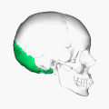 Position of occipital bone (shown in green). Animation.
 -
 Outer surface
 -
 Inner surface. Frontal bone and parietal bones are removed.
 -
 Occipital bone
 -
 Occipital bone
 -
 Median sagittal section through the occipital bone and first three cervical vertebræ
 -
 Basilar part
 -
 Occipital bone


See also
- Cerebellum
- Neanderthal
- Occipital bun
- Occipital lobe
- Ossification of occipital bone
References
Books
![]() This article incorporates text in the public domain from page 129 of the 20th edition of Gray's Anatomy (1918)
This article incorporates text in the public domain from page 129 of the 20th edition of Gray's Anatomy (1918)
- Susan Standring; Neil R. Borley; et al., eds. (2008). Gray's anatomy: the anatomical basis of clinical practice (40th ed.). London: Churchill Livingstone. ISBN 978-0-8089-2371-8.
Citations
- S2CID 1091809– via Taylor & Francis Online.
- ^ Gray's Anatomy 2008, p. 424-425.
- ^ Gray's Anatomy 2008, p. 425.
- ^ Hacking, Craig. "Basion-dens interval | Radiology Reference Article | Radiopaedia.org". radiopaedia.org. Retrieved 5 December 2016.
- ^ "occipital" A Dictionary of Zoology. Ed. Michael Allaby. Oxford University Press 2009
- ^ ISBN 0-03-910284-X.
External links
 Media related to Occipital bones at Wikimedia Commons